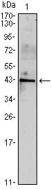
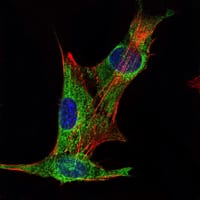
INHA Primary Antibody
Inhibins are peptide hormones produced by the granulosa cells in female follicles and by Sertoli cells in the male seminiferous tubules. They are selectively expressed by cells of sex cord stromal derivation, and inhibit the secretion of follitropin by the pituitary gland. Inhibins are also involved in regulating diverse functions such as hypothalamic and pituitary hormone secretion, gonadal hormone secretion, germ cell development and maturation, erythroid differentiation, insulin secretion, nerve cell survival, embryonic axial development or bone growth, depending on their subunit composition. Inhibins appear to oppose the functions of activins, as inhibins and activins inhibit and activate, respectively, the secretion of follitropin by the pituitary gland. Inhibin has 2 subunits (alpha and beta) that are coded by separate genes. The alpha subunit determines whether inhibin or activin will be produced. The alpha subunit remains constant, such that the various types of inhibin are defined by the beta subunit (a,b,c,d). Inhibin A is a dimer of alpha and beta A. Inhibin B is a dimer of alpha and beta B. Proteolytic processing yields a number of inhibin alpha bioactive forms: the 20/23 kDa forms consist solely of the mature alpha chain, the 26/29 kDa forms consist of the most N terminal propeptide linked through a disulfide bond to the mature alpha chain, and the 50/53 kDa forms encompass the entire proprotein. Each type can be furthermore either mono or diglycosylated, causing the mass difference.
2. Acta Histochem. 2009;111(4):360-5.
3. Hum Reprod. 2009 Aug;24(8):2023-8.