STAT3 Primary Antibody

Item Information
Catalog #
Size
Price
Description

The protein encoded by this gene is a member of the STAT protein family. In response to cytokines and growth factors, STAT family members are phosphorylated by the receptor associated kinases, and then form homo- or heterodimers that translocate to the cell nucleus where they act as transcription activators. This protein is activated through phosphorylation in response to various cytokines and growth factors including IFNs, EGF, IL5, IL6, HGF, LIF and BMP2. This protein mediates the expression of a variety of genes in response to cell stimuli, and thus plays a key role in many cellular processes such as cell growth and apoptosis. The small GTPase Rac1 has been shown to bind and regulate the activity of this protein. PIAS3 protein is a specific inhibitor of this protein. Three alternatively spliced transcript variants encoding distinct isoforms have been described.

Product Overview
Entrez GenelD
6774
Aliases
APRF; HIES; FLJ20882; MGC16063; STAT3
Host / Isotype
Rabbit / IgG
Species Reactivity
Human, Mouse, Rat
Immunogen
Synthesized non-phosphopeptide derived from human STAT3 around the phosphorylation site of serine 727.
Formulation
The antibody was affinity-purified from rabbit antiserum by affinity-chromatography using epitope-specific immunogen. Liquid in PBS containing 50% glycerol and 0.03% sodium azide.
Storage
4°C; -20°C for long term storage
Product Applications
WB (Western Blot)
1/500 - 1/2000
IHC_P(Immunohistochemistry)
1/200 - 1/1000
ICC (Immunocytochemistry)
1/200 - 1/1000
ELISA
1/10000
References
1. J Biol Chem. 2010 Jul 16;285(29):22414-25.
2. Int J Oncol. 2010 Jul;37(1):103-10.
3. Clin Immunol. 2010 Sep;136(3):442-6.
Product Image
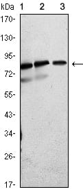
Western Blot
Figure 1: Western blot analysis using STAT3 Rabbit pAb against Jurkat (1), Hela (2) and PC-12 (3) cell lysate.
Immunofluorescence analysis
Figure 2:Immunofluorescence analysis of SMMC-7721 cells using STAT3 Rabbit pAb (Red). Blue: DRAQ5 fluorescent DNA dye.
Immunohistochemical analysis
igure 3: Immunohistochemical analysis of paraffin-embedded ovary tumor (left), recta cancer tissues (right) using STAT3 Rabbit pAb with DAB staining.
For Research Use Only. Not for use in diagnostic procedures.