

PKHD1 Primary Antibody
Item Information
Catalog #
Size
Price
Description
Mutations in the PKHD1 gene, which encodes fibrocystin, cause autosomal recessive polycystic kidney disease (ARPKD). Unfortunately, the lack of specific antibodies to the mouse protein impairs the study of splicing, post-translational processing, shedding, and temporal and spatial expression of endogenous fibrocystin at the cellular and subcellular level.
Product Overview
Entrez GenelD
241035
Aliases
FPC; Tigm1; AI118496; AI182499
Clone#
8G12A1
Host / Isotype
Rat / IgG1
Species Reactivity
Human
Immunogen
Purified recombinant fragment of mouse PKHD1 (AA: 3878-4060) expressed in E. Coli.
Formulation
Purified antibody from tissue culture in PBS with 0.05% sodium azide
Storage
Store at 4°C short term. Aliquot and store at -20°C long term. Avoid freeze/thaw cycles.
Product Applications
IHC_P(Immunohistochemistry)
1/200 - 1/1000
ICC (Immunocytochemistry)
1/200 - 1/1000
FCM (Flow Cytometry)
1/200 - 1/400
ELISA
1/10000
References
J Am Soc Nephrol. 2011 Dec;22(12):2266-77.
Am J Pathol. 2008 Feb;172(2):417-29.
Am J Pathol. 2008 Feb;172(2):417-29.
Product Image
Western Blot
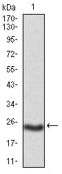
Figure 1: Western blot analysis using PKHD1 mAb against mouse PKHD1(AA: 3878-4060) recombinant protein. (Expected MW is 23 kDa)
Immunofluorescence analysis
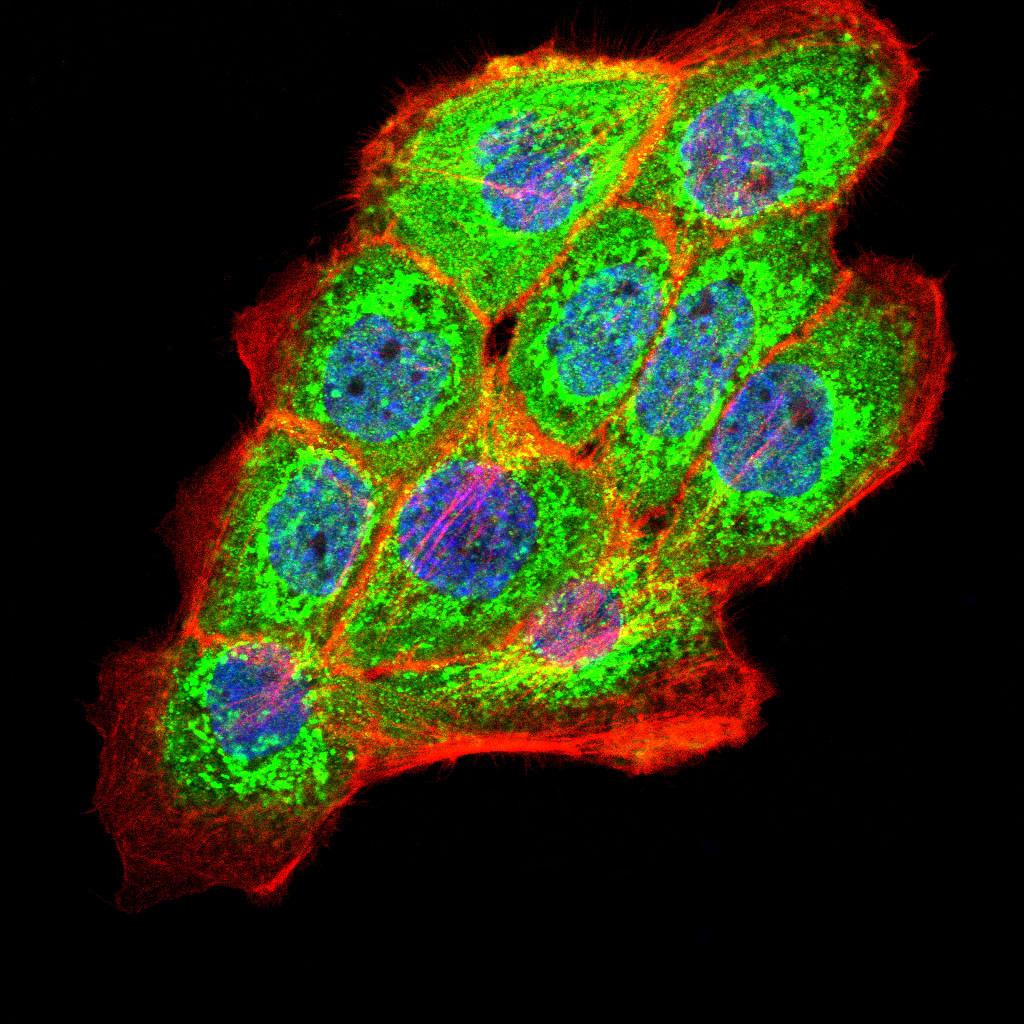
Figure 2:Immunofluorescence analysis of A431 cells using PKHD1 Rat mAb (green). Blue: DRAQ5 fluorescent DNA dye. Red: Actin filaments have been labeled with Alexa Fluor- 555 phalloidin. Secondary antibody from Fisher (Cat#: 35503)
Flow cytometric
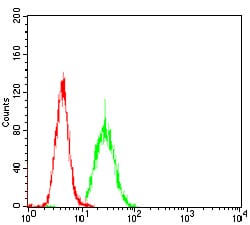
Figure 3: Flow cytometric analysis of Hela cells using PKHD1 Rat mAb (green) and negative control (red).
Immunohistochemical analysis
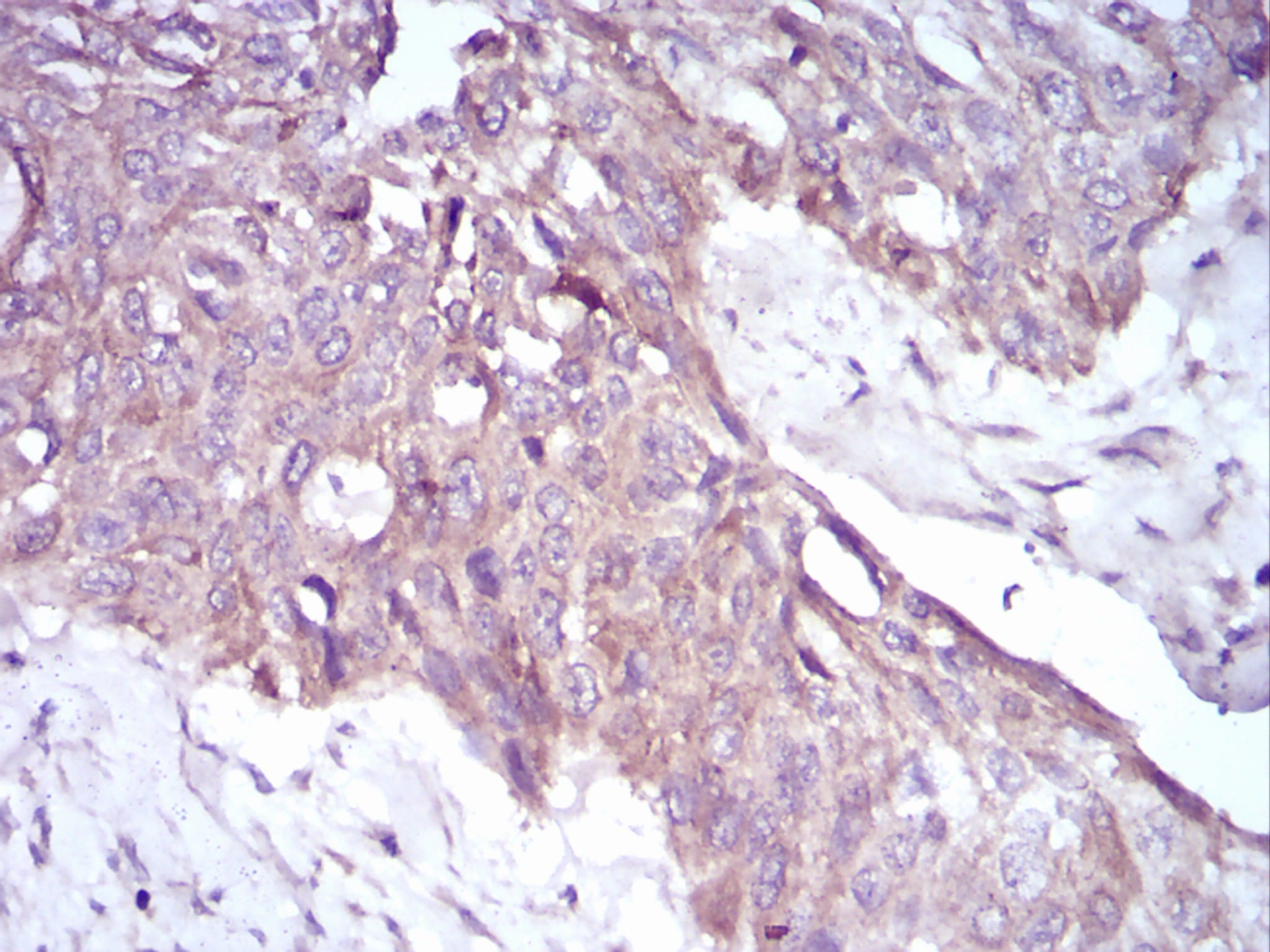
Figure 4: Immunohistochemical analysis of paraffin-embedded esophageal cancer tissues using PKHD1 Rat mAb with DAB staining.
Elisa
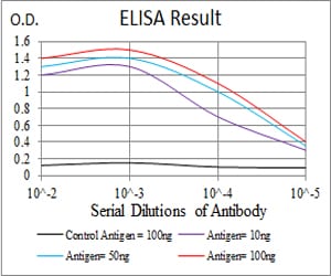
Black line: Control Antigen (100 ng); Purple line: Antigen(10ng); Blue line: Antigen (50 ng); Red line: Antigen (100 ng);
For Research Use Only. Not for use in diagnostic procedures.