

ZP2 Primary Antibody
Item Information
Catalog #
Size
Price
Description
The zona pellucida is an extracellular matrix that surrounds the oocyte and early embryo. It is composed of three glycoproteins with various functions during fertilization and preimplantation development. The glycosylated mature peptide is one of the structural components of the zona pellucida and functions in secondary binding and penetration of acrosome-reacted spermatozoa. Female mice lacking this gene do not form a stable zona matrix and are sterile. Alternative splicing results in multiple transcript variants.
Product Overview
Entrez GenelD
7783
Aliases
ZPA; Zp-2; OOMD6
Clone#
2F11E4
Host / Isotype
Mouse / Mouse IgG1
Species Reactivity
Human, Mouse, Rat
Immunogen
Purified recombinant fragment of human ZP2 (AA: 624-745) expressed in E. Coli.
Formulation
Purified antibody in PBS with 0.05% sodium azide
Storage
Store at 4°C short term. Aliquot and store at -20°C long term. Avoid freeze/thaw cycles.
Product Applications
WB (Western Blot)
1/500 - 1/2000
IHC_P(Immunohistochemistry)
1/200 - 1/1000
FCM (Flow Cytometry)
1/200 - 1/400
ELISA
1/10000
References
1.J Assist Reprod Genet. 2021 May;38(5):1239-1245.
2.J Assist Reprod Genet. 2020 Nov;37(11):2853-2860.
2.J Assist Reprod Genet. 2020 Nov;37(11):2853-2860.
Product Image
Elisa

Figure 1:Black line: Control Antigen (100 ng);Purple line: Antigen (10ng); Blue line: Antigen (50 ng); Red line:Antigen (100 ng)
Western Blot
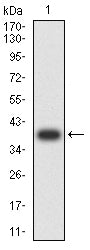
Figure 2:Western blot analysis using ZP2 mAb against human ZP2 (AA: 624-745) recombinant protein. (Expected MW is 38.5 kDa)
Western Blot

Figure 3:Western blot analysis using ZP2 mAb against HEK293-6e (1) and ZP2 (AA: 624-745)-hIgGFc transfected HEK293-6e (2) cell lysate.
Immunofluorescence analysis

Figure 4:Flow cytometric analysis of Hela cells using ZP2 mouse mAb (green) and negative control (red).
Immunohistochemical analysis

Figure 5:Immunohistochemical analysis of paraffin-embedded breast cancer tissues using ZP2 mouse mAb with DAB staining.
Immunohistochemical analysis

Figure 6:Immunohistochemical analysis of paraffin-embedded colon cancer tissues using ZP2 mouse mAb with DAB staining.
Immunohistochemical analysis

Figure 7:Immunohistochemical analysis of paraffin-embedded rectum cancer tissues using ZP2 mouse mAb with DAB staining.
Immunohistochemical analysis

Figure 8:Immunohistochemical analysis of paraffin-embedded mouse kidney tissues using ZP2 mouse mAb with DAB staining.
Immunohistochemical analysis

Figure 9:Immunohistochemical analysis of paraffin-embedded Rat kidney tissues using ZP2 mouse mAb with DAB staining.
Immunohistochemical analysis

Figure 10:Immunohistochemical analysis of paraffin-embedded Rat cerebrum tissues using ZP2 mouse mAb with DAB staining.
Immunohistochemical analysis

Figure 11:Immunohistochemical analysis of paraffin-embedded Rabbit kidney tissues using ZP2 mouse mAb with DAB staining.
Immunohistochemical analysis

Figure 12:Immunohistochemical analysis of paraffin-embedded Rabbit spinal cord tissues using ZP2 mouse mAb with DAB staining.
For Research Use Only. Not for use in diagnostic procedures.