

ZEB1 Primary Antibody
Item Information
Catalog #
Size
Price
Description
This gene encodes a zinc finger transcription factor. The encoded protein likely plays a role in transcriptional repression of interleukin 2. Mutations in this gene have been associated with posterior polymorphous corneal dystrophy-3 and late-onset Fuchs endothelial corneal dystrophy. Alternatively spliced transcript variants encoding different isoforms have been described.
Product Overview
Entrez GenelD
6935
Aliases
BZP; TCF8; AREB6; FECD6; NIL2A; PPCD3; ZFHEP; ZFHX1A; DELTAEF1
Clone#
2A8H3
Host / Isotype
Mouse / IgG1
Species Reactivity
Human
Immunogen
Purified recombinant fragment of human ZEB1 (AA: 967-1108) expressed in E. Coli.
Formulation
Purified antibody in PBS with 0.05% sodium azide
Storage
Store at 4°C short term. Aliquot and store at -20°C long term. Avoid freeze/thaw cycles.
Product Applications
WB (Western Blot)
1/500 - 1/2000
IHC_P(Immunohistochemistry)
1/200 - 1/1000
ICC (Immunocytochemistry)
1/200 - 1/1000
FCM (Flow Cytometry)
1/200 - 1/400
ELISA
1/10000
References
1. J Cancer Res Clin Oncol. 2012 Aug;138(8):1329-38.
2. Mol Cell Biochem. 2012 Jul;366(1-2):223-9.
2. Mol Cell Biochem. 2012 Jul;366(1-2):223-9.
Product Image
Western Blot

Figure 1: Western blot analysis using ZEB1 mAb against human ZEB1 recombinant protein. (Expected MW is 41.7 kDa)
Western Blot

Figure 2: Western blot analysis using ZEB1 mAb against HEK293 (1) and ZEB1 (AA: 967-1108)-hIgGFc transfected HEK293 (2) cell lysate.
Immunofluorescence analysis

Figure 3: Immunofluorescence analysis of Hela cells using ZEB1 mouse mAb (green). Blue: DRAQ5 fluorescent DNA dye. Red: Actin filaments have been labeled with Alexa Fluor-555 phalloidin.
Flow cytometric

Figure 4: Flow cytometric analysis of Hela cells using ZEB1 mouse mAb (green) and negative control (red).
Immunohistochemical analysis
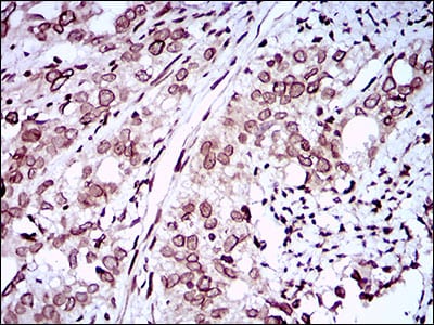
Figure 5: Immunohistochemical analysis of paraffin-embedded cervical cancer tissues using ZEB1 mouse mAb with DAB staining.
Immunohistochemical analysis

Figure 6: Immunohistochemical analysis of paraffin-embedded rectum cancer tissues using ZEB1 mouse mAb with DAB staining.
Elisa

Black line: Control Antigen (100 ng); Purple line: Antigen(10ng); Blue line: Antigen (50 ng); Red line: Antigen (100 ng);
For Research Use Only. Not for use in diagnostic procedures.