

XRN2 Primary Antibody
Item Information
Catalog #
Size
Price
Description
This gene encodes a 5'-3' exonuclease that promotes transcription termination at cotranscriptional cleavage sites. Alternative splicing results in multiple transcript variants encoding different isoforms.
Product Overview
Entrez GenelD
22803
Aliases
N
Clone#
9F7G11
Host / Isotype
Mouse / IgG1
Species Reactivity
Human
Immunogen
Purified recombinant fragment of human XRN2 (AA: 398-547) expressed in E. Coli.
Formulation
Purified antibody in PBS with 0.05% sodium azide
Storage
Store at 4°C short term. Aliquot and store at -20°C long term. Avoid freeze/thaw cycles.
Product Applications
WB (Western Blot)
1/500 - 1/2000
IHC_P(Immunohistochemistry)
1/200 - 1/1000
ELISA
1/10000
References
1.EMBO J. 2012 May 30;31(11):2566-78.
2.DNA Seq. 2005 Apr;16(2):143-6.
2.DNA Seq. 2005 Apr;16(2):143-6.
Product Image
Elisa
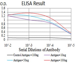
Figure 1: Black line: Control Antigen (100 ng);Purple line: Antigen (10ng); Blue line: Antigen (50 ng); Red line:Antigen (100 ng)
Western Blot

Figure 2:Western blot analysis using XRN2 mAb against human XRN2 (AA: 398-547) recombinant protein. (Expected MW is 43.1 kDa)
Western Blot

Figure 3:Western blot analysis using XRN2 mAb against HEK293 (1) and XRN2 (AA: 398-547)-hIgGFc transfected HEK293 (2) cell lysate.
Western Blot

Figure 4:Western blot analysis using XRN2 mouse mAb against HEK293 (1), NTERA-2 (2), LNcap (3), HepG2 (4), and PC-3 (5) cell lysate.
Immunohistochemical analysis

Figure 5:Immunohistochemical analysis of paraffin-embedded esophageal cancer tissues using XRN2 mouse mAb with DAB staining.
Immunohistochemical analysis

Figure 6:Immunohistochemical analysis of paraffin-embedded rectum cancer tissues using XRN2 mouse mAb with DAB staining.
For Research Use Only. Not for use in diagnostic procedures.