
XRCC6 Primary Antibody
Item Information
Catalog #
Size
Price
Description
The p70/p80 autoantigen is a nuclear complex consisting of two subunits with molecular masses of approximately 70 and 80 kDa. The complex functions as a single-stranded DNA-dependent ATP-dependent helicase. The complex may be involved in the repair of nonhomologous DNA ends such as that required for double-strand break repair, transposition, and V(D)J recombination. High levels of autoantibodies to p70 and p80 have been found in some patients with systemic lupus erythematosus.
Product Overview
Entrez GenelD
2547
Aliases
ML8; KU70; TLAA; CTC75; CTCBF; G22P1
Clone#
7A9E7
Host / Isotype
Mouse / IgG1
Species Reactivity
Human
Immunogen
Purified recombinant fragment of human XRCC6 (AA: 6-214) expressed in E. Coli.
Formulation
Purified antibody in PBS with 0.05% sodium azide.
Storage
Store at 4°C short term. Aliquot and store at -20°C long term. Avoid freeze/thaw cycles.
Product Applications
WB (Western Blot)
1/500 - 1/2000
ICC (Immunocytochemistry)
1/200 - 1/1000
FCM (Flow Cytometry)
1/200 - 1/400
ELISA
1/10000
References
1. Clin Cancer Res. 2013 Mar 15;19(6):1547-56.
2. Mol Carcinog. 2012 Oct;51 Suppl 1:E183-90.
2. Mol Carcinog. 2012 Oct;51 Suppl 1:E183-90.
Product Image
Western Blot

Figure 1: Western blot analysis using XRCC6 mAb against human XRCC6 (AA: 6-214) recombinant protein. (Expected MW is 49.7 kDa)
Western Blot

Figure 2: Western blot analysis using XRCC6 mAb against HEK293 (1) and XRCC6 (AA: 6-214)-hIgGFc transfected HEK293 (2) cell lysate.
Western Blot

Figure 3: Western blot analysis using XRCC6 mouse mAb against PC-2 (1), A549 (2), A431 (3), HepG2 (4), K562 (5) cell lysate.
Immunofluorescence analysis

Figure 4: Immunofluorescence analysis of MCF-7 cells using XRCC6 mouse mAb (green). Blue: DRAQ5 fluorescent DNA dye. Red: Actin filaments have been labeled with Alexa Fluor-555 phalloidin. Secondary antibody from Fisher (Cat#: 35503)
Flow cytometric
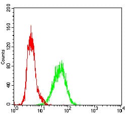
Figure 5: Flow cytometric analysis of A431 cells using XRCC6 mouse mAb (green) and negative control (red).
Elisa

Black line: Control Antigen (100 ng); Purple line: Antigen(10ng); Blue line: Antigen (50 ng); Red line: Antigen (100 ng);
For Research Use Only. Not for use in diagnostic procedures.