

XPC Primary Antibody
Item Information
Catalog #
Size
Price
Description
The protein encoded by this gene is a key component of the XPC complex, which plays an important role in the early steps of global genome nucleotide excision repair (NER). The encoded protein is important for damage sensing and DNA binding, and shows a preference for single-stranded DNA. Mutations in this gene or some other NER components can result in Xeroderma pigmentosum, a rare autosomal recessive disorder characterized by increased sensitivity to sunlight with the development of carcinomas at an early age. Alternatively spliced transcript variants have been found for this gene.
Product Overview
Entrez GenelD
7508
Aliases
XP3; RAD4; XPCC; p125
Clone#
5F4D3
Host / Isotype
Mouse / Mouse IgG1
Species Reactivity
Human
Immunogen
Purified recombinant fragment of human XPC (AA: 32-133) expressed in mammalian.
Formulation
Purified antibody in PBS with 0.05% sodium azide
Storage
Store at 4°C short term. Aliquot and store at -20°C long term. Avoid freeze/thaw cycles.
Product Applications
WB (Western Blot)
1/500 - 1/2000
IHC_P(Immunohistochemistry)
1/200 - 1/1000
ICC (Immunocytochemistry)
1/50 - 1/200
FCM (Flow Cytometry)
1/200 - 1/400
ELISA
1/10000
References
1,Acta Medica (Hradec Kralove). 2020;63(3):101-112.
2,Nat Commun. 2020 Nov 17;11(1):5834.
2,Nat Commun. 2020 Nov 17;11(1):5834.
Product Image
Elisa

Figure 1:Black line: Control Antigen (100 ng);Purple line: Antigen (10ng); Blue line: Antigen (50 ng); Red line:Antigen (100 ng)
Western Blot

Figure 2:Western blot analysis using XPC mAb against human XPC (AA: 32-133) recombinant protein. (Expected MW is 41.9 kDa)
Western Blot
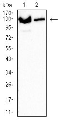
Figure 3:Western blot analysis using XPC mouse mAb against Jurkat (1) and Hela (2) cell lysate.
Immunohistochemical analysis

Figure 4:Immunofluorescence analysis of Hela cells using XPC mouse mAb (green). Blue: DRAQ5 fluorescent DNA dye. Red: Actin filaments have been labeled with Alexa Fluor- 555 phalloidin. Secondary antibody from Fisher (Cat#: 35503)
Immunofluorescence analysis
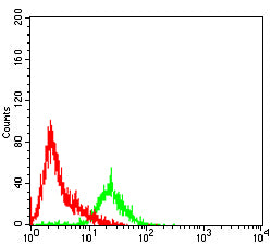
Figure 5:Flow cytometric analysis of Hela cells using XPC mouse mAb (green) and negative control (red).
Immunohistochemical analysis
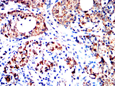
Figure 6:Immunohistochemical analysis of paraffin-embedded cervical cancer tissues using XPC mouse mAb with DAB staining.
Immunohistochemical analysis

Figure 7:Immunohistochemical analysis of paraffin-embedded ovarian cancer tissues using XPC mouse mAb with DAB staining.
For Research Use Only. Not for use in diagnostic procedures.