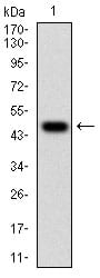
WT1 Primary Antibody

Item Information
Catalog #
Size
Price
Description

This gene encodes a transcription factor that contains four zinc-finger motifs at the C-terminus and a proline/glutamine-rich DNA-binding domain at the N-terminus. It has an essential role in the normal development of the urogenital system, and it is mutated in a small subset of patients with Wilm's tumors. This gene exhibits complex tissue-specific and polymorphic imprinting pattern, with biallelic, and monoallelic expression from the maternal and paternal alleles in different tissues. Multiple transcript variants have been described. In several variants, there is evidence for the use of a non-AUG (CUG) translation initiation site upstream of and in-frame with the first AUG. Authors of PMID:7926762 also provide evidence that WT1 mRNA undergoes RNA editing in human and rat, and that this process is tissue-restricted and developmentally regulated.

Product Overview
Entrez GenelD
7490
Aliases
GUD; AWT1; WAGR; WT33; NPHS4; WIT-2; EWS-WT1
Clone#
5G11A5
Host / Isotype
Mouse / IgG1
Species Reactivity
Human
Immunogen
Purified recombinant fragment of human WT1 (AA: 314-479) expressed in E. Coli.
Formulation
Purified antibody from tissue culture in PBS with 0.05% sodium azide
Storage
4°C; -20°C for long term storage
Product Applications
WB (Western Blot)
1/500 - 1/2000
FCM (Flow Cytometry)
1/200 - 1/400
ELISA
1/10000
References
1. Leuk Res. 2013 Oct;37(10):1341-9.
2. Pediatr Blood Cancer. 2013 Aug;60(8):1388-9.
Product Image
Western Blot
Figure 1: Western blot analysis using WT1 mAb against human WT1 (AA: 314-479) recombinant protein. (Expected MW is 47.6 kDa)
Western Blot
Figure 2: Western blot analysis using WT1 mAb against HEK293 (1) and WT1 (AA: 314-479)-hIgGFc transfected HEK293 (2) cell lysate.
Flow cytometric
Figure 3: Flow cytometric analysis of Hela cells using WT1 mouse mAb (green) and negative control (red).
Elisa
Black line: Control Antigen (100 ng); Purple line: Antigen(10ng); Blue line: Antigen (50 ng); Red line: Antigen (100 ng);
For Research Use Only. Not for use in diagnostic procedures.