
WNT5A Primary Antibody
Item Information
Catalog #
Size
Price
Description
WNT5A: wingless-type MMTV integration site family, member 5A. Entrez Protein: NP_003383. The WNT gene family consists of structurally related genes which encode secreted signaling proteins. These proteins have been implicated in oncogenesis and in several developmental processes, including regulation of cell fate and patterning during embryogenesis. This gene is a member of the WNT gene family. It encodes a protein which shows 98%, 98% and 87% amino acid identity to the mouse, rat and the xenopus Wnt5A protein, respectively. The experiments performed in Xenopus laevis embryos identified that human frizzled-5 (hFz5) is the receptor for the Wnt5A ligand and the Wnt5A/hFz5 signaling mediates axis induction.
Product Overview
Entrez GenelD
7474
Aliases
hWNT5A
Clone#
6F2
Host / Isotype
Mouse / IgG1
Species Reactivity
Human
Immunogen
Purified recombinant fragment of WNT5A expressed in E. Coli.
Formulation
Ascitic fluid containing 0.03% sodium azide.
Storage
Store at 4°C short term. Aliquot and store at -20°C long term. Avoid freeze/thaw cycles.
Product Applications
WB (Western Blot)
1/500 - 1/2000
IHC_P(Immunohistochemistry)
1/200 - 1/1000
ICC (Immunocytochemistry)
1/200 - 1/1000
ELISA
1/10000
References
1. Cancer Res. 2008 Jul 15;68(14):5785-94.
2. Proc Natl Acad Sci U S A. 2009 Mar 10;106(10):3919-24.
2. Proc Natl Acad Sci U S A. 2009 Mar 10;106(10):3919-24.
Product Image
Western Blot

Figure 1: Western blot analysis using WNT5A mouse mAb against HEK293 (1) and WNT5A-hIgGFc transfected HEK293 cell lysate (2).
Immunohistochemical analysis
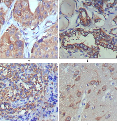
Figure 2: Immunohistochemical analysis of paraffin-embedded human lung cancer (A), thyroid cancer (B), lymph node (C) and brain (D) showing cytoplasmic and extracellular matrix localization using WNT5A mouse mAb with DAB staining.
Immunofluorescence analysis

Figure 3: Confocal Immunofluorescence analysis of PC-12 cells using WNT5A mouse mAb (green), showing cytoplasmic localization. Blue: DRAQ5 fluorescent DNA dye.
For Research Use Only. Not for use in diagnostic procedures.