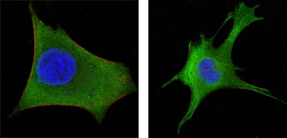
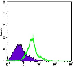
WNT1 Primary Antibody

Item Information
Catalog #
Size
Price
Description

WNT1: wingless-type MMTV integration site family, member 1. The WNT gene family consists of structurally related genes which encode secreted signaling proteins. These proteins have been implicated in oncogenesis and in several developmental processes, including regulation of cell fate and patterning during embryogenesis. This gene is a member of the WNT gene family. It is very conserved in evolution, and the protein encoded by this gene is known to be 98% identical to the mouse Wnt1 protein at the amino acid level. The studies in mouse indicate that the Wnt1 protein functions in the induction of the mesencephalon and cerebellum. This gene was originally considered as a candidate gene for Joubert syndrome, an autosomal recessive disorder with cerebellar hypoplasia as a leading feature. However, further studies suggested that the gene mutations might not have a significant rolein Joubert syndrome. This gene is clustered with another family member, WNT10B, in the chromosome 12q13 region.

Product Overview
Entrez GenelD
7471
Aliases
INT1
Clone#
10C8
Host / Isotype
Mouse / IgG1
Species Reactivity
Human, Mouse
Immunogen
Purified recombinant fragment of WNT1 expressed in E. Coli.
Formulation
Ascitic fluid containing 0.03% sodium azide.
Storage
4°C; -20°C for long term storage
Product Applications
WB (Western Blot)
1/500 - 1/2000
IHC_P(Immunohistochemistry)
1/200 - 1/1000
ICC (Immunocytochemistry)
1/200 - 1/1000
FCM (Flow Cytometry)
1/200 - 1/400
ELISA
1/10000
References
1. Blood. 2008 Jan 1;111(1):122-31.
2. BMC Cancer. 2005 May 24;5:53.
Product Image
Western Blot
Figure 1: Western blot analysis using WNT1 mouse mAb against NIH/3T3 (1), 3T3L1 (2) and Hela (3) cell lysate.
Immunohistochemical analysis
Figure 2: Immunohistochemical analysis of paraffin-embedded human LAdrenal tissues using WNT1 mouse mAb
Immunofluorescence analysis
Figure3: Confocal Immunofluorescence analysis of Hela (left) and 3T3-L1 (right) cells using WNT1 mouse mAb (green). Red: Actin filaments have been labeled with DY-554 phalloidin. Blue: DRAQ5 fluorescent DNA dye.
Flow cytometric
Figure 4: Flow cytometric analysis of Hela cells using WNT1 mouse mAb (green) and negative control (purple).
For Research Use Only. Not for use in diagnostic procedures.