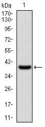
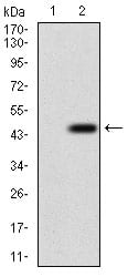
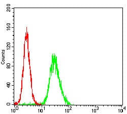
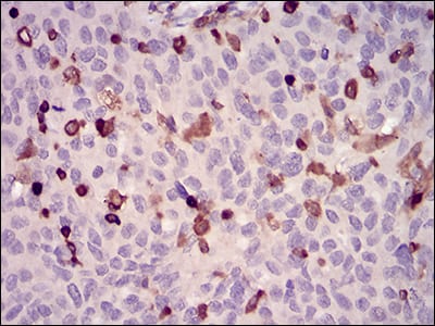
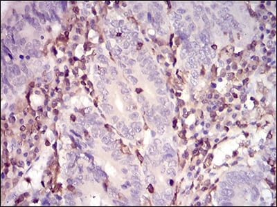
WAS Primary Antibody

Item Information
Catalog #
Size
Price
Description

The Wiskott-Aldrich syndrome (WAS) family of proteins share similar domain structure, and are involved in transduction of signals from receptors on the cell surface to the actin cytoskeleton. The presence of a number of different motifs suggests that they are regulated by a number of different stimuli, and interact with multiple proteins. Recent studies have demonstrated that these proteins, directly or indirectly, associate with the small GTPase, Cdc42, known to regulate formation of actin filaments, and the cytoskeletal organizing complex, Arp2/3. Wiskott-Aldrich syndrome is a rare, inherited, X-linked, recessive disease characterized by immune dysregulation and microthrombocytopenia, and is caused by mutations in the WAS gene. The WAS gene product is a cytoplasmic protein, expressed exclusively in hematopoietic cells, which show signalling and cytoskeletal abnormalities in WAS patients. A transcript variant arising as a result of alternative promoter usage, and containing a different 5' UTR sequence, has been described, however, its full-length nature is not known.

Product Overview
Entrez GenelD
7454
Aliases
THC; IMD2; SCNX; THC1; WASP
Clone#
7B10E4
Host / Isotype
Mouse / IgG2a
Species Reactivity
Human
Immunogen
Purified recombinant fragment of human WAS (AA: 57-170) expressed in E. Coli.
Formulation
Purified antibody from tissue culture in PBS with 0.05% sodium azide
Storage
4°C; -20°C for long term storage
Product Applications
WB (Western Blot)
1/500 - 1/2000
IHC_P(Immunohistochemistry)
1/200 - 1/1000
FCM (Flow Cytometry)
1/200 - 1/400
ELISA
1/10000
References
1. Mol Cell Biol. 2012 Aug;32(15):3153-63.
2. Dis Markers. 2010;29(3-4):157-75.
Product Image
Western Blot
Figure 1: Western blot analysis using WAS mAb against human WAS (AA: 57-170) recombinant protein. (Expected MW is 39 kDa)
Western Blot
Figure 2: Western blot analysis using WAS mAb against HEK293 (1) and WAS (AA: 57-170)-hIgGFc transfected HEK293 (2) cell lysate.
Flow cytometric
Figure 3: Flow cytometric analysis of Hela cells using WAS mouse mAb (green) and negative control (red).
Immunohistochemical analysis
Figure 4: Immunohistochemical analysis of paraffin-embedded ovarian cancer tissues using WAS mouse mAb with DAB staining.
Immunohistochemical analysis
Figure 5: Immunohistochemical analysis of paraffin-embedded colon cancer tissues using WAS mouse mAb with DAB staining.
Elisa
Black line: Control Antigen (100 ng); Purple line: Antigen(10ng); Blue line: Antigen (50 ng); Red line: Antigen (100 ng);
For Research Use Only. Not for use in diagnostic procedures.