

VTN Primary Antibody
Item Information
Catalog #
Size
Price
Description
The protein encoded by this gene is a member of the pexin family. It is found in serum and tissues and promotes cell adhesion and spreading, inhibits the membrane-damaging effect of the terminal cytolytic complement pathway, and binds to several serpin serine protease inhibitors. It is a secreted protein and exists in either a single chain form or a clipped, two chain form held together by a disulfide bond.
Product Overview
Entrez GenelD
7448
Aliases
VN; V75; VNT
Clone#
1G11E8
Host / Isotype
Mouse / IgG2b
Species Reactivity
Human
Immunogen
Purified recombinant fragment of human VTN (AA: 20-199) expressed in E. Coli.
Formulation
Purified antibody from tissue culture in PBS with 0.05% sodium azide
Storage
Store at 4°C short term. Aliquot and store at -20°C long term. Avoid freeze/thaw cycles.
Product Applications
WB (Western Blot)
1/500 - 1/2000
IHC_P(Immunohistochemistry)
1/200 - 1/1000
FCM (Flow Cytometry)
1/200 - 1/400
ELISA
1/10000
References
1. Inflamm Res. 2012 Nov;61(11):1241-6.
2. J Cancer Res Clin Oncol. 2011 Jul;137(7):1105-15.
2. J Cancer Res Clin Oncol. 2011 Jul;137(7):1105-15.
Product Image
Western Blot
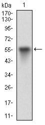
Figure 1: Western blot analysis using VTN mAb against human VTN (AA: 20-199) recombinant protein. (Expected MW is 45.9 kDa)
Western Blot
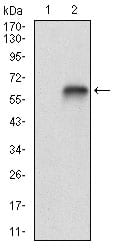
Figure 2: Western blot analysis using VTN mAb against HEK293 (1) and VTN (AA: 20-199)-hIgGFc transfected HEK293 (2) cell lysate.
Flow cytometric
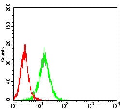
Figure 3: Flow cytometric analysis of Hela cells using VTN mouse mAb (green) and negative control (red).
Immunohistochemical analysis
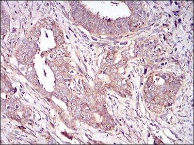
Figure 4: Immunohistochemical analysis of paraffin-embedded cervical cancer tissues using VTN mouse mAb with DAB staining.
Immunohistochemical analysis
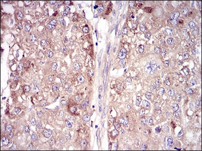
Figure 5: Immunohistochemical analysis of paraffin-embedded liver cancer tissues using VTN mouse mAb with DAB staining.
Elisa
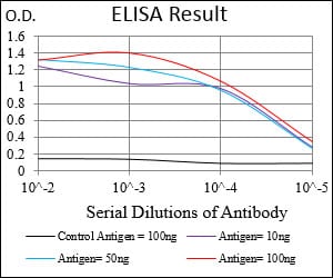
Black line: Control Antigen (100 ng); Purple line: Antigen(10ng); Blue line: Antigen (50 ng); Red line: Antigen (100 ng);
For Research Use Only. Not for use in diagnostic procedures.