

VIMP Primary Antibody
Item Information
Catalog #
Size
Price
Description
This gene encodes a member of the selenoprotein family, characterized by a selenocysteine (Sec) residue at the active site. The selenocysteine is encoded by the UGA codon that normally signals translation termination. The 3' UTR of selenoprotein genes have a common stem-loop structure, the sec insertion sequence (SECIS), that is necessary for the recognition of UGA as a Sec codon rather than as a stop signal. Studies suggest that this protein may regulate cytokine production, and thus play a key role in the control of the inflammatory response. Alternative splicing results in multiple transcript variants encoding different isoforms.
Product Overview
Entrez GenelD
55829
Aliases
SELS; ADO15; SBBI8; SEPS1; AD-015
Clone#
5G4A10
Host / Isotype
Mouse / IgG1
Species Reactivity
Human
Immunogen
Purified recombinant fragment of human VIMP (AA: 1-187) expressed in E. Coli.
Formulation
Purified antibody in PBS with 0.05% sodium azide
Storage
Store at 4°C short term. Aliquot and store at -20°C long term. Avoid freeze/thaw cycles.
Product Applications
WB (Western Blot)
1/500 - 1/2000
ICC (Immunocytochemistry)
1/50 - 1/250
FCM (Flow Cytometry)
1/200 - 1/400
ELISA
1/10000
References
1.Free Radic Biol Med. 2014 Feb;67:265-77.
2.PLoS One. 2013 Jun 11;8(6):e65657.
2.PLoS One. 2013 Jun 11;8(6):e65657.
Product Image
Elisa
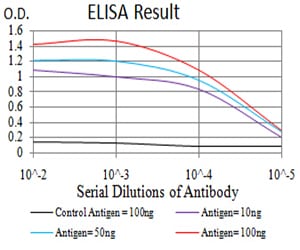
Figure 1: Black line: Control Antigen (100 ng);Purple line: Antigen (10ng); Blue line: Antigen (50 ng); Red line:Antigen (100 ng)
Western Blot
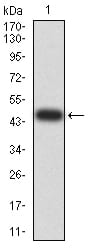
Figure 2:Western blot analysis using VIMP mAb against human VIMP (AA: 1-187) recombinant protein. (Expected MW is 46.9 kDa)
Western Blot
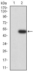
Figure 3:Western blot analysis using VIMP mAb against HEK293 (1) and VIMP (AA: 1-187)-hIgGFc transfected HEK293 (2) cell lysate.
Immunofluorescence analysis
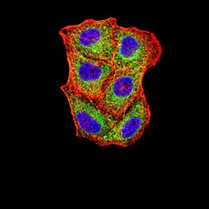
Figure 4:Immunofluorescence analysis of Hela cells using VIMP mouse mAb (green). Blue: DRAQ5 fluorescent DNA dye. Red: Actin filaments have been labeled with Alexa Fluor- 555 phalloidin. Secondary antibody from Fisher (Cat#: 35503)
Flow cytometric
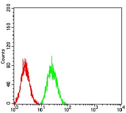
Figure 5:Flow cytometric analysis of Hela cells using VIMP mouse mAb (green) and negative control (red).
For Research Use Only. Not for use in diagnostic procedures.