

VIL1 Primary Antibody
Item Information
Catalog #
Size
Price
Description
This gene encodes a member of a family of calcium-regulated actin-binding proteins. This protein represents a dominant part of the brush border cytoskeleton which functions in the capping, severing, and bundling of actin filaments. Two mRNAs of 2.7 kb and 3.5 kb have been observed; they result from utilization of alternate poly-adenylation signals present in the terminal exon.
Product Overview
Entrez GenelD
7429
Aliases
VIL; D2S1471
Clone#
3E5G11
Host / Isotype
Mouse / IgG1
Species Reactivity
Human
Immunogen
Purified recombinant fragment of human VIL1 (AA: 1-209) expressed in E. Coli.
Formulation
Purified antibody in PBS with 0.05% sodium azide.
Storage
Store at 4°C short term. Aliquot and store at -20°C long term. Avoid freeze/thaw cycles.
Product Applications
WB (Western Blot)
1/500 - 1/2000
IHC_P(Immunohistochemistry)
1/200 - 1/1000
ICC (Immunocytochemistry)
1/200 - 1/1000
FCM (Flow Cytometry)
1/200 - 1/400
ELISA
1/10000
References
1. Cancer Sci. 2012 Aug;103(8):1493-501.
2. Cancer Biol Ther. 2011 Aug 1;12(3):181-90.
2. Cancer Biol Ther. 2011 Aug 1;12(3):181-90.
Product Image
Western Blot

Figure 1: Western blot analysis using VIL1 mAb against human VIL1 (AA: 1-209) recombinant protein. (Expected MW is 49.4 kDa)
Western Blot
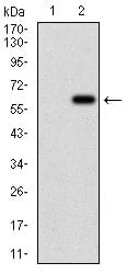
Figure 2: Western blot analysis using VIL1 mAb against HEK293 (1) and VIL1 (AA: 1-209)-hIgGFc transfected HEK293 (2) cell lysate.
Western Blot
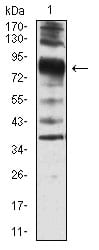
Figure 3: Western blot analysis using VIL1 mouse mAb against SW620 cell lysate.
Immunofluorescence analysis

Figure 4:Immunofluorescence analysis of Hela cells using VIL1 mouse mAb (green). Blue: DRAQ5 fluorescent DNA dye. Red: Actin filaments have been labeled with Alexa Fluor- 555 phalloidin. Secondary antibody from Fisher (Cat#: 35503)
Flow cytometric
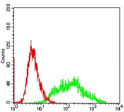
Figure 5: Flow cytometric analysis of SW620 cells using VIL1 mouse mAb (green) and negative control (red).
Immunohistochemical analysis

Figure 6: Immunohistochemical analysis of paraffin-embedded cervical cancer tissues using VIL1 mouse mAb with DAB staining.
Elisa

Black line: Control Antigen (100 ng); Purple line: Antigen(10ng); Blue line: Antigen (50 ng); Red line: Antigen (100 ng);
For Research Use Only. Not for use in diagnostic procedures.