

VIL1 Primary Antibody
Item Information
Catalog #
Size
Price
Description
This gene encodes a member of a family of calcium-regulated actin-binding proteins. This protein represents a dominant part of the brush border cytoskeleton which functions in the capping, severing, and bundling of actin filaments. Two mRNAs of 2.7 kb and 3.5 kb have been observed; they result from utilization of alternate poly-adenylation signals present in the terminal exon.
Product Overview
Entrez GenelD
7429
Aliases
VIL; D2S1471
Clone#
5E3B2
Host / Isotype
Mouse / IgG2b
Species Reactivity
Human
Immunogen
Purified recombinant fragment of human VIL1 (AA: 1-209) expressed in E. Coli.
Formulation
Purified antibody from tissue culture in PBS with 0.05% sodium azide
Storage
Store at 4°C short term. Aliquot and store at -20°C long term. Avoid freeze/thaw cycles.
Product Applications
WB (Western Blot)
1/500 - 1/2000
IHC_P(Immunohistochemistry)
1/200 - 1/1000
ELISA
1/10000
References
1. Cancer Biol Ther. 2011 Aug 1;12(3):181-90.
2. Cancer Biol Ther. 2009 Jun;8(12):1146-53.
2. Cancer Biol Ther. 2009 Jun;8(12):1146-53.
Product Image
Western Blot

Figure 1: Western blot analysis using VIL1 mAb against human VIL1 (AA: 1-209) recombinant protein. (Expected MW is 49.4 kDa)
Western Blot

Figure 2: Western blot analysis using VIL1 mAb against HEK293 (1) and VIL1 (AA: 1-209)-hIgGFc transfected HEK293 (2) cell lysate.
Immunohistochemical analysis
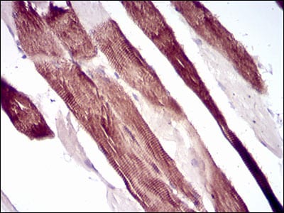
Figure 3: Immunohistochemical analysis of paraffin-embedded muscle tissues using VIL1 mouse mAb with DAB staining.
Immunohistochemical analysis

Figure 4: Immunohistochemical analysis of paraffin-embedded stomach cancer tissues using VIL1 mouse mAb with DAB staining.
Elisa

Black line: Control Antigen (100 ng); Purple line: Antigen(10ng); Blue line: Antigen (50 ng); Red line: Antigen (100 ng);
For Research Use Only. Not for use in diagnostic procedures.