

VEGFA Primary Antibody
Item Information
Catalog #
Size
Price
Description
This gene is a member of the PDGF/VEGF growth factor family. It encodes a heparin-binding protein, which exists as a disulfide-linked homodimer. This growth factor induces proliferation and migration of vascular endothelial cells, and is essential for both physiological and pathological angiogenesis. Disruption of this gene in mice resulted in abnormal embryonic blood vessel formation. This gene is upregulated in many known tumors and its expression is correlated with tumor stage and progression. Elevated levels of this protein are found in patients with POEMS syndrome, also known as Crow-Fukase syndrome. Allelic variants of this gene have been associated with microvascular complications of diabetes 1 (MVCD1) and atherosclerosis. Alternatively spliced transcript variants encoding different isoforms have been described. There is also evidence for alternative translation initiation from upstream non-AUG (CUG) codons resulting in additional isoforms. A recent study showed that a C-terminally extended isoform is produced by use of an alternative in-frame translation termination codon via a stop codon readthrough mechanism, and that this isoform is antiangiogenic. Expression of some isoforms derived from the AUG start codon is regulated by a small upstream open reading frame, which is located within an internal ribosome entry site.
Product Overview
Entrez GenelD
7422
Aliases
VPF; VEGF; MVCD1
Clone#
6G5A10
Host / Isotype
Mouse / IgG1
Species Reactivity
Human
Immunogen
Purified recombinant fragment of human VEGFA (AA: 207-371) expressed in E. Coli.
Formulation
Purified antibody in PBS with 0.05% sodium azide
Storage
Store at 4°C short term. Aliquot and store at -20°C long term. Avoid freeze/thaw cycles.
Product Applications
WB (Western Blot)
1/500 - 1/2000
FCM (Flow Cytometry)
1/200 - 1/400
ELISA
1/10000
References
1.Oncol Rep. 2014 Dec;32(6):2359-64.
2.Eur J Cancer. 2014 Nov;50(16):2855-65.
2.Eur J Cancer. 2014 Nov;50(16):2855-65.
Product Image
Elisa

Figure 1: Black line: Control Antigen (100 ng);Purple line: Antigen (10ng); Blue line: Antigen (50 ng); Red line:Antigen (100 ng)
Western Blot

Figure 2:Western blot analysis using VEGFA mAb against human VEGFA (AA: 207-371) recombinant protein. (Expected MW is 20.6 kDa)
Western Blot
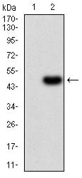
Figure 3:Western blot analysis using VEGFA mAb against HEK293 (1) and VEGFA (AA: 207-371)-hIgGFc transfected HEK293 (2) cell lysate.
Western Blot
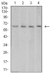
Figure 4:Western blot analysis using VEGFA mouse mAb against HUVEC (1), HEK293 (2), Jurkat (3), and Hela (4) cell lysate.
Flow cytometric
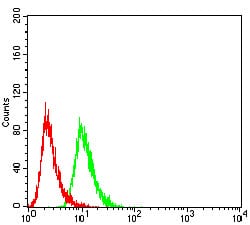
Figure 5:Flow cytometric analysis of Hela cells using VEGFA mouse mAb (green) and negative control (red).
For Research Use Only. Not for use in diagnostic procedures.