

UPK3B Primary Antibody
Item Information
Catalog #
Size
Price
Description
UPK3B is a minor component of the apical plaques of mammalian urothelium that binds and dimerizes with uroplakin-1b (UPK1B; MIM 602380), one of the major conserved urothelium membrane proteins. The other major conserved integral membrane proteins of urothelial plaques are UPK1A (MIM 611557), UPK2 (MIM 611558), and UPK3A (MIM 611559) (Deng et al., 2002 [PubMed 12446744]).
Product Overview
Entrez GenelD
105375355
Aliases
P35; UP3B; UPIIIB
Clone#
3B2G1
Host / Isotype
Mouse / Mouse IgG1
Species Reactivity
Human
Immunogen
Purified recombinant fragment of human UPK3B (AA: 30-180) expressed in E. Coli.
Formulation
Purified antibody in PBS with 0.05% sodium azide
Storage
Store at 4°C short term. Aliquot and store at -20°C long term. Avoid freeze/thaw cycles.
Product Applications
WB (Western Blot)
1/500 - 1/2000
ICC (Immunocytochemistry)
1/50 - 1/200
FCM (Flow Cytometry)
1/200 - 1/400
ELISA
1/10000
References
1.PLoS One. 2011;6(10):e25391.
2.Mol Phylogenet Evol. 2006 Nov;41(2):355-67.
2.Mol Phylogenet Evol. 2006 Nov;41(2):355-67.
Product Image
Elisa
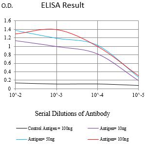
Figure 1:Black line: Control Antigen (100 ng);Purple line: Antigen (10ng); Blue line: Antigen (50 ng); Red line:Antigen (100 ng)
Western Blot

Figure 2:Western blot analysis using UPK3B mAb against human UPK3B (AA: 30-180) recombinant protein. (Expected MW is 41.5 kDa)
Western Blot

Figure 3:Western blot analysis using UPK3B mAb against HEK293-6e (1) and UPK3B (AA: 30-180)-hIgGFc transfected HEK293-6e (2) cell lysate.
Immunohistochemical analysis

Figure 4:Immunofluorescence analysis of Hela cells using UPK3B mouse mAb (green). Blue: DRAQ5 fluorescent DNA dye. Red: Actin filaments have been labeled with Alexa Fluor- 555 phalloidin. Secondary antibody from Fisher (Cat#: 35503)
Immunofluorescence analysis
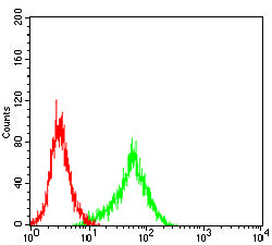
Figure 5:Flow cytometric analysis of THP-1 cells using UPK3B mouse mAb (green) and negative control (red).
For Research Use Only. Not for use in diagnostic procedures.