

ULK2 Primary Antibody
Item Information
Catalog #
Size
Price
Description
This gene encodes a protein that is similar to a serine/threonine kinase in C. elegans which is involved in axonal elongation. The structure of this protein is similar to the C. elegans protein in that both proteins have an N-terminal kinase domain, a central proline/serine rich (PS) domain, and a C-terminal (C) domain. The gene is located within the Smith-Magenis syndrome region on chromosome 17. Alternatively spliced transcript variants encoding the same protein have been identified.
Product Overview
Entrez GenelD
9706
Aliases
ATG1B; Unc51.2
Clone#
2H4B2
Host / Isotype
Mouse / IgG1
Species Reactivity
Human, Mouse, Rat
Immunogen
Purified recombinant fragment of human ULK2 (AA: 1-155) expressed in E. Coli.
Formulation
Purified antibody in PBS with 0.05% sodium azide
Storage
Store at 4°C short term. Aliquot and store at -20°C long term. Avoid freeze/thaw cycles.
Product Applications
WB (Western Blot)
1/500 - 1/2000
FCM (Flow Cytometry)
1/200 - 1/400
ELISA
1/10000
References
1.J Biol Chem. 2014 Aug 8;289(32):22306-18.
2.Oncogene. 1999 Oct 21;18(43):5850-9.
2.Oncogene. 1999 Oct 21;18(43):5850-9.
Product Image
Elisa
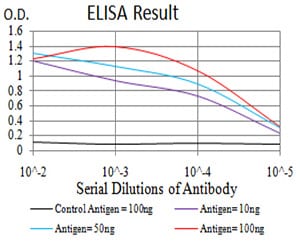
Figure 1: Black line: Control Antigen (100 ng);Purple line: Antigen (10ng); Blue line: Antigen (50 ng); Red line:Antigen (100 ng)
Western Blot
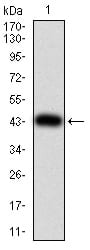
Figure 2:Western blot analysis using ULK2 mAb against human ULK2 (AA: 1-155) recombinant protein. (Expected MW is 43.4 kDa)
Western Blot
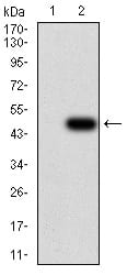
Figure 3:Western blot analysis using ULK2 mAb against HEK293 (1) and ULK2 (AA: 1-155)-hIgGFc transfected HEK293 (2) cell lysate.
Western Blot
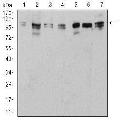
Figure 4:Western blot analysis using ULK2 mouse mAb against NIH/3T3 (1), HepG2 (2), SK-Hep-1 (3), SK-OV-3 (4), C6 (5), PC-12 (6), and MCF-7 (7) cell lysate.
Flow cytometric
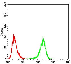
Figure 5:Flow cytometric analysis of Hela cells using ULK2 mouse mAb (green) and negative control (red).
For Research Use Only. Not for use in diagnostic procedures.