

ULBP1 Primary Antibody
Item Information
Catalog #
Size
Price
Description
The protein encoded by this gene is a ligand of natural killer group 2, member D (NKG2D), an immune system-activating receptor on NK cells and T-cells. Binding of the encoded ligand to NKG2D leads to activation of several signal transduction pathways, including those of JAK2, STAT5, ERK and PI3K kinase/Akt. Also, in cytomegalovirus-infected cells, this ligand binds the UL16 glycoprotein and is prevented from activating the immune system. Three transcript variants encoding different isoforms have been found for this gene. [provided by RefSeq, Nov 2015]
Product Overview
Entrez GenelD
80329
Aliases
N2DL-1; RAET1I; NKG2DL1
Clone#
3A6F11
Host / Isotype
Mouse / Mouse IgG1
Immunogen
Purified recombinant fragment of human ULBP1 (AA: 26-216) expressed in E. Coli.
Formulation
Purified antibody in PBS with 0.05% sodium azide
Storage
Store at 4°C short term. Aliquot and store at -20°C long term. Avoid freeze/thaw cycles.
Product Applications
WB (Western Blot)
1/500 - 1/2000
FCM (Flow Cytometry)
1/200 - 1/400
ELISA
1/10000
References
1.Oncotarget. 2016 Mar 29;7(13):15369-81. 2.Elife. 2015 Nov 13;4. pii: e08474.
Product Image
Elisa
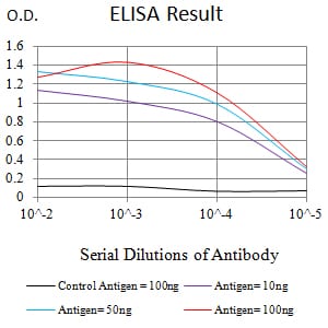
Figure 1:Black line: Control Antigen (100 ng);Purple line: Antigen (10ng); Blue line: Antigen (50 ng); Red line:Antigen (100 ng)
Western Blot
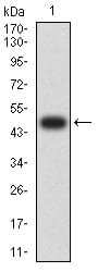
Figure 2:Western blot analysis using ULBP1 mAb against human ULBP1 (AA: 26-216) recombinant protein. (Expected MW is 48.3 kDa)
Western Blot
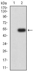
Figure 3:Western blot analysis using ULBP1 mAb against HEK293 (1) and ULBP1 (AA: 26-216)-hIgGFc transfected HEK293 (2) cell lysate.
Flow cytometric
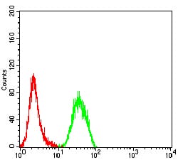
Figure 4:Flow cytometric analysis of Hela cells using ULBP1 mouse mAb (green) and negative control (red).
For Research Use Only. Not for use in diagnostic procedures.