

UFD1L Primary Antibody
Item Information
Catalog #
Size
Price
Description
The protein encoded by this gene forms a complex with two other proteins, nuclear protein localization-4 and valosin-containing protein, and this complex is necessary for the degradation of ubiquitinated proteins. In addition, this complex controls the disassembly of the mitotic spindle and the formation of a closed nuclear envelope after mitosis. Mutations in this gene have been associated with Catch 22 syndrome as well as cardiac and craniofacial defects. Alternative splicing results in multiple transcript variants encoding different isoforms. A related pseudogene has been identified on chromosome 18.
Product Overview
Entrez GenelD
7353
Aliases
UFD1
Clone#
2A6F3
Host / Isotype
Mouse / IgG2b
Species Reactivity
Human
Immunogen
Purified recombinant fragment of human UFD1L (AA: 208-307) expressed in E. Coli.
Formulation
Purified antibody in PBS with 0.05% sodium azide
Storage
Store at 4°C short term. Aliquot and store at -20°C long term. Avoid freeze/thaw cycles.
Product Applications
WB (Western Blot)
1/500 - 1/2000
IHC_P(Immunohistochemistry)
1/200 - 1/1000
ICC (Immunocytochemistry)
1/200 - 1/1000
FCM (Flow Cytometry)
1/200 - 1/400
ELISA
1/10000
References
1.Proc Natl Acad Sci U S A. 2011 May 31;108(22):9119-24.
2.Cell Biochem Funct. 2003 Sep;21(3):263-7.V
2.Cell Biochem Funct. 2003 Sep;21(3):263-7.V
Product Image
Elisa
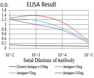
Figure 1: Black line: Control Antigen (100 ng);Purple line: Antigen (10ng); Blue line: Antigen (50 ng); Red line:Antigen (100 ng)
Western Blot

Figure 2:Western blot analysis using UFD1L mAb against human UFD1L (AA: 208-307) recombinant protein. (Expected MW is 36.8 kDa)
Western Blot
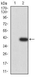
Figure 3:Western blot analysis using UFD1L mAb against HEK293 (1) and UFD1L (AA: 208-307)-hIgGFc transfected HEK293 (2) cell lysate.
Western Blot
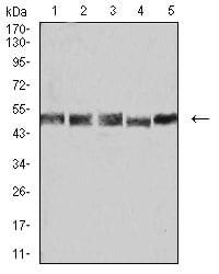
Figure 4:Western blot analysis using UFD1L mouse mAb against K562 (1), Hela (2), A431 (3), PC-2 (4), and A549 (5) cell lysate.
Immunofluorescence analysis
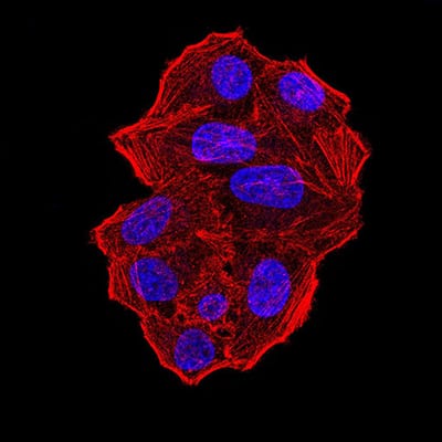
Figure 5:Immunofluorescence analysis of Hela cells using UFD1L mouse mAb. Blue: DRAQ5 fluorescent DNA dye. Red: Actin filaments have been labeled with Alexa Fluor- 555 phalloidin.
Immunofluorescence analysis
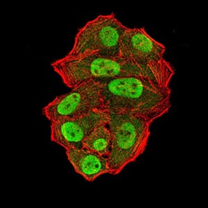
Figure 6:Immunofluorescence analysis of Hela cells using UFD1L mouse mAb (green). Blue: DRAQ5 fluorescent DNA dye. Red: Actin filaments have been labeled with Alexa Fluor- 555 phalloidin. Secondary antibody from Fisher (Cat#: 35503)
Flow cytometric
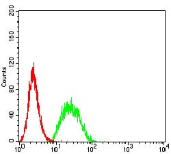
Figure 7:Flow cytometric analysis of Hela cells using UFD1L mouse mAb (green) and negative control (red).
Immunohistochemical analysis
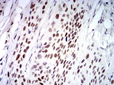
Figure 8:Immunohistochemical analysis of paraffin-embedded esophageal cancer tissues using UFD1L mouse mAb with DAB staining.
Immunohistochemical analysis
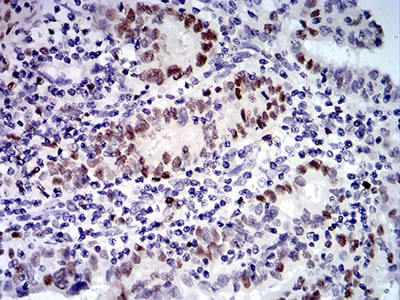
Figure 9:Immunohistochemical analysis of paraffin-embedded endometrial cancer tissues using UFD1L mouse mAb with DAB staining.
For Research Use Only. Not for use in diagnostic procedures.