

UCP3 Primary Antibody
Item Information
Catalog #
Size
Price
Description
Mitochondrial uncoupling proteins (UCP) are members of the larger family of mitochondrial anion carrier proteins (MACP). UCPs separate oxidative phosphorylation from ATP synthesis with energy dissipated as heat, also referred to as the mitochondrial proton leak. UCPs facilitate the transfer of anions from the inner to the outer mitochondrial membrane and the return transfer of protons from the outer to the inner mitochondrial membrane. They also reduce the mitochondrial membrane potential in mammalian cells. The different UCPs have tissue-specific expression; this gene is primarily expressed in skeletal muscle. This gene's protein product is postulated to protect mitochondria against lipid-induced oxidative stress. Expression levels of this gene increase when fatty acid supplies to mitochondria exceed their oxidation capacity and the protein enables the export of fatty acids from mitochondria. UCPs contain the three solcar protein domains typically found in MACPs. Two splice variants have been found for this gene.
Product Overview
Entrez GenelD
7352
Aliases
SLC25A9
Clone#
6B8C6
Host / Isotype
Mouse / IgG2a
Species Reactivity
Human
Immunogen
Purified recombinant fragment of human UCP3 (AA: 1-113 and 217-312) expressed in E. Coli.
Formulation
Purified antibody in PBS with 0.05% sodium azide
Storage
Store at 4°C short term. Aliquot and store at -20°C long term. Avoid freeze/thaw cycles.
Product Applications
WB (Western Blot)
1/500 - 1/2000
ICC (Immunocytochemistry)
1/200 - 1/1000
ELISA
1/10000
References
1.J Biol Chem. 2011 Sep 16;286(37):32533-41.
2.Nutr Hosp. 2012 Jul-Aug;27(4):1190-5.
2.Nutr Hosp. 2012 Jul-Aug;27(4):1190-5.
Product Image
Elisa
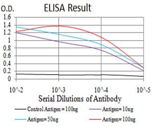
Figure 1: Black line: Control Antigen (100 ng);Purple line: Antigen (10ng); Blue line: Antigen (50 ng); Red line:Antigen (100 ng)
Western Blot
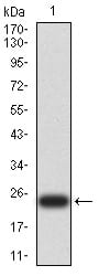
Figure 2:Western blot analysis using UCP3 mAb against human UCP3 (AA: 1-113 and 217-312) recombinant protein. (Expected MW is 24 kDa)
Western Blot
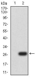
Figure 3:Western blot analysis using UCP3 mAb against HEK293 (1) and UCP3 (AA:1-113 and 217-312)-hIgGFc transfected HEK293 (2) cell lysate.
Immunofluorescence analysis
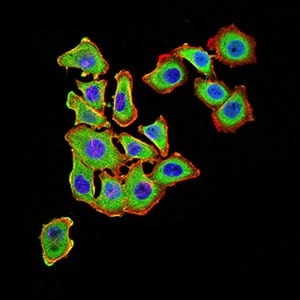
Figure 4:Immunofluorescence analysis of HL-7702 cells using UCP3 mouse mAb (green). Blue: DRAQ5 fluorescent DNA dye. Red: Actin filaments have been labeled with Alexa Fluor- 555 phalloidin. Secondary antibody from Fisher (Cat#: 35503)
For Research Use Only. Not for use in diagnostic procedures.