

UCP2 Primary Antibody
Item Information
Catalog #
Size
Price
Description
Mitochondrial uncoupling proteins (UCP) are members of the larger family of mitochondrial anion carrier proteins (MACP). UCPs separate oxidative phosphorylation from ATP synthesis with energy dissipated as heat, also referred to as the mitochondrial proton leak. UCPs facilitate the transfer of anions from the inner to the outer mitochondrial membrane and the return transfer of protons from the outer to the inner mitochondrial membrane. They also reduce the mitochondrial membrane potential in mammalian cells. Tissue specificity occurs for the different UCPs and the exact methods of how UCPs transfer H+/OH- are not known. UCPs contain the three homologous protein domains of MACPs. This gene is expressed in many tissues, with the greatest expression in skeletal muscle. It is thought to play a role in nonshivering thermogenesis, obesity and diabetes. Chromosomal order is 5'-UCP3-UCP2-3'.
Product Overview
Entrez GenelD
7351
Aliases
UCPH; BMIQ4; SLC25A8
Clone#
3F1B9
Host / Isotype
Mouse / IgG2a
Species Reactivity
Human
Immunogen
Purified recombinant fragment of human UCP2 (AA: 1-309) expressed in E. Coli.
Formulation
Purified antibody in PBS with 0.05% sodium azide
Storage
Store at 4°C short term. Aliquot and store at -20°C long term. Avoid freeze/thaw cycles.
Product Applications
WB (Western Blot)
1/500 - 1/2000
ICC (Immunocytochemistry)
1/200 - 1/1000
FCM (Flow Cytometry)
1/200 - 1/400
ELISA
1/10000
References
1.Endocrine. 2013 Jun;43(3):714-23.
2.Carcinogenesis. 2012 Nov;33(11):2065-75.
2.Carcinogenesis. 2012 Nov;33(11):2065-75.
Product Image
Elisa

Figure 1: Black line: Control Antigen (100 ng);Purple line: Antigen (10ng); Blue line: Antigen (50 ng); Red line:Antigen (100 ng)
Western Blot

Figure 2:Western blot analysis using UCP2 mAb against human UCP2 (AA: 1-309) recombinant protein. (Expected MW is 36.1 kDa)
Immunofluorescence analysis

Figure 4:Immunofluorescence analysis of Hela cells using UCP2 mouse mAb (green). Blue: DRAQ5 fluorescent DNA dye. Red: Actin filaments have been labeled with Alexa Fluor- 555 phalloidin. Secondary antibody from Fisher (Cat#: 35503)
Flow cytometric

Figure 5:Flow cytometric analysis of Hela cells using UCP2 mouse mAb (green) and negative control (red).
Western Blot
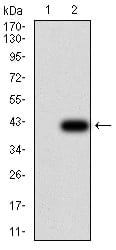
Figure 5:Western blot analysis using UCP2 mAb against HEK293 (1) and UCP2 (AA: 1-309)-hIgGFc transfected HEK293 (2) cell lysate.
For Research Use Only. Not for use in diagnostic procedures.