

UCHL1 Primary Antibody
Item Information
Catalog #
Size
Price
Description
The protein encoded by this gene belongs to the peptidase C12 family. This enzyme is a thiol protease that hydrolyzes a peptide bond at the C-terminal glycine of ubiquitin. This gene is specifically expressed in the neurons and in cells of the diffuse neuroendocrine system. Mutations in this gene may be associated with Parkinson disease.
Product Overview
Entrez GenelD
7345
Aliases
NDGOA; PARK5; PGP95; SPG79; PGP9.5; UCHL-1; Uch-L1; HEL-117; PGP 9.5; HEL-S-53
Clone#
1D1B12
Host / Isotype
Mouse / Mouse IgG2b
Species Reactivity
Human, Mouse, Rat
Immunogen
Purified recombinant fragment of human UCHL1 (AA: 1-220) expressed in E. Coli.
Formulation
Purified antibody in PBS with 0.05% sodium azide
Storage
Store at 4°C short term. Aliquot and store at -20°C long term. Avoid freeze/thaw cycles.
Product Applications
WB (Western Blot)
1/500 - 1/2000
IHC_P(Immunohistochemistry)
1/200 - 1/1000
FCM (Flow Cytometry)
1/200 - 1/400
ELISA
1/10000
References
1.Endocr Relat Cancer. 2019 Apr 1;26(4):411-423.
2.Cancer Sci. 2020 Feb;111(2):610-620.
2.Cancer Sci. 2020 Feb;111(2):610-620.
Product Image
Elisa

Figure 1:Black line: Control Antigen (100 ng);Purple line: Antigen (10ng); Blue line: Antigen (50 ng); Red line:Antigen (100 ng)
Western Blot

Figure 2:Western blot analysis using UCHL1 mAb against human UCHL1 (AA: 1-220) recombinant protein. (Expected MW is 27.5 kDa)
Western Blot

Figure 3:Western blot analysis using UCHL1 mAb against HEK293 (1) and UCHL1 (AA: 1-220)-hIgGFc transfected HEK293 (2) cell lysate.
Western Blot

Figure 4:Western blot analysis using UCHL1 mouse mAb against DU145 (1), A549 (2) cell lysate, rat brain (3), and mouse brain (4) tissue lysate.
Immunofluorescence analysis
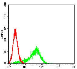
Figure 5:Flow cytometric analysis of Hela cells using UCHL1 mouse mAb (green) and negative control (red).
Immunohistochemical analysis

Figure 6:Immunohistochemical analysis of paraffin-embedded bladder cancer tissues using UCHL1 mouse mAb with DAB staining.
Immunohistochemical analysis

Figure 7:Immunohistochemical analysis of paraffin-embedded lung cancer tissues using UCHL1 mouse mAb with DAB staining.
Immunohistochemical analysis

Figure 8:Immunohistochemical analysis of paraffin-embedded cervical cancer tissues using UCHL1 mouse mAb with DAB staining.
Immunohistochemical analysis

Figure 9:Immunohistochemical analysis of paraffin-embedded esophageal cancer tissues using UCHL1 mouse mAb with DAB staining.
Immunohistochemical analysis

Figure 10:Immunohistochemical analysis of paraffin-embedded mouse brain tissues using UCHL1 mouse mAb with DAB staining.
Immunohistochemical analysis

Figure 11:Immunohistochemical analysis of paraffin-embedded mouse kidney tissues using UCHL1 mouse mAb with DAB staining.
Immunohistochemical analysis

Figure 12:Immunohistochemical analysis of paraffin-embedded rat brain tissues using UCHL1 mouse mAb with DAB staining.
Immunohistochemical analysis

Figure 13:Immunohistochemical analysis of paraffin-embedded rat kidney tissues using UCHL1 mouse mAb with DAB staining.
For Research Use Only. Not for use in diagnostic procedures.