

UBE2C Primary Antibody
Item Information
Catalog #
Size
Price
Description
The modification of proteins with ubiquitin is an important cellular mechanism for targeting abnormal or short-lived proteins for degradation. Ubiquitination involves at least three classes of enzymes: ubiquitin-activating enzymes, ubiquitin-conjugating enzymes, and ubiquitin-protein ligases. This gene encodes a member of the E2 ubiquitin-conjugating enzyme family. The encoded protein is required for the destruction of mitotic cyclins and for cell cycle progression, and may be involved in cancer progression. Multiple transcript variants encoding different isoforms have been found for this gene. Pseudogenes of this gene have been defined on chromosomes 4, 14, 15, 18, and 19.
Product Overview
Entrez GenelD
11065
Aliases
UBCH10; dJ447F3.2
Clone#
1F5D3
Host / Isotype
Mouse / IgG2a
Species Reactivity
Human
Immunogen
Purified recombinant fragment of human UBE2C (AA: FULL(1-179)) expressed in E. Coli.
Formulation
Purified antibody from tissue culture in PBS with 0.05% sodium azide
Storage
4°C; -20°C for long term storage
Product Applications
WB (Western Blot)
1/500 - 1/2000
IHC_P(Immunohistochemistry)
1/200 - 1/1000
ELISA
1/10000
References
1. J Cancer Res Clin Oncol. 2012 Nov;138(11):1951-61.
2. Histopathology. 2009 May;54(6):731-40.
2. Histopathology. 2009 May;54(6):731-40.
Product Image
Western Blot
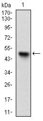
Figure 1: Western blot analysis using UBE2C mAb against human UBE2C (AA: FULL(1-179)) recombinant protein. (Expected MW is 45.6 kDa)
Western Blot
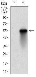
Figure 2: Western blot analysis using UBE2C mAb against HEK293 (1) and UBE2C (AA: FULL(1-179))-hIgGFc transfected HEK293 (2) cell lysate.
Immunohistochemical analysis
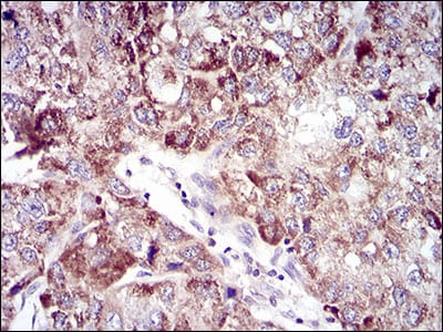
Figure 3: Immunohistochemical analysis of paraffin-embedded liver cancer tissues using UBE2C mouse mAb with DAB staining.
Immunohistochemical analysis
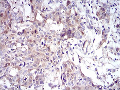
Figure 4: Immunohistochemical analysis of paraffin-embedded esophageal cancer tissues using UBE2C mouse mAb with DAB staining.
Western Blot
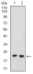
Figure 6:Western blot analysis using UBE2C mouse mAb against Hela (1), and Raji (2) cell lysate.
Elisa
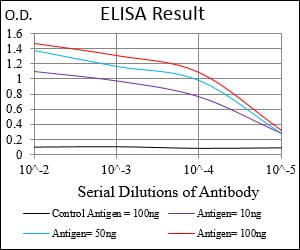
Black line: Control Antigen (100 ng); Purple line: Antigen(10ng); Blue line: Antigen (50 ng); Red line: Antigen (100 ng);
For Research Use Only. Not for use in diagnostic procedures.