

TYRO3 Primary Antibody
Item Information
Catalog #
Size
Price
Description
The gene is part of a 3-member transmembrane receptor kinase receptor family with a processed pseudogene distal on chromosome 15. The encoded protein is activated by the products of the growth arrest-specific gene 6 and protein S genes and is involved in controlling cell survival and proliferation, spermatogenesis, immunoregulation and phagocytosis. The encoded protein has also been identified as a cell entry factor for Ebola and Marburg viruses.
Product Overview
Entrez GenelD
7301
Aliases
BYK; Dtk; RSE; Rek; Sky; Tif; Etk-2
Clone#
2F5B8
Host / Isotype
Mouse / Mouse IgG1
Species Reactivity
Human
Immunogen
Purified recombinant fragment of human TYRO3 (AA: extra 230-429) expressed in mammalian.
Formulation
Purified antibody in PBS with 0.05% sodium azide
Storage
Store at 4°C short term. Aliquot and store at -20°C long term. Avoid freeze/thaw cycles.
Product Applications
WB (Western Blot)
1/500 - 1/2000
IHC_P(Immunohistochemistry)
1/200 - 1/1000
FCM (Flow Cytometry)
1/200 - 1/400
ELISA
1/10000
References
1,Anticancer Res. 2020 Oct;40(10):5593-5600.
2,Anticancer Res. 2020 Nov;40(11):6115-6121.
2,Anticancer Res. 2020 Nov;40(11):6115-6121.
Product Image
Elisa

Figure 1:Black line: Control Antigen (100 ng);Purple line: Antigen (10ng); Blue line: Antigen (50 ng); Red line:Antigen (100 ng)
Western Blot

Figure 2:Western blot analysis using TYRO3 mAb against human TYRO3 (AA: 230-429) recombinant protein. (Expected MW is 52 kDa)
Immunofluorescence analysis

Figure 3:Flow cytometric analysis of U937 cells using TYRO3 mouse mAb (green) and negative control (red).
Immunohistochemical analysis
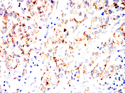
Figure 4:Immunohistochemical analysis of paraffin-embedded liver cancer tissues using TYRO3 mouse mAb with DAB staining.
Immunohistochemical analysis

Figure 5:Immunohistochemical analysis of paraffin-embedded human brain tissues using TYRO3 mouse mAb with DAB staining.
For Research Use Only. Not for use in diagnostic procedures.