

TWIST1 Primary Antibody
Item Information
Catalog #
Size
Price
Description
Basic helix-loop-helix (bHLH) transcription factors have been implicated in cell lineage determination and differentiation. The protein encoded by this gene is a bHLH transcription factor and shares similarity with another bHLH transcription factor, Dermo1. The strongest expression of this mRNA is in placental tissue; in adults, mesodermally derived tissues express this mRNA preferentially. Mutations in this gene have been found in patients with Saethre-Chotzen syndrome.
Product Overview
Entrez GenelD
7291
Aliases
SCS; ACS3; CRS1; BPES2; BPES3; TWIST; bHLHa38
Clone#
10E4E6
Host / Isotype
Mouse / IgG1
Species Reactivity
Human, Mouse
Immunogen
Purified recombinant fragment of human TWIST1 (AA: 9-74) expressed in E. Coli.
Formulation
Purified antibody in PBS with 0.05% sodium azide
Storage
Store at 4°C short term. Aliquot and store at -20°C long term. Avoid freeze/thaw cycles.
Product Applications
WB (Western Blot)
1/500 - 1/2000
IHC_P(Immunohistochemistry)
1/200 - 1/1000
ICC (Immunocytochemistry)
1/200 - 1/1000
FCM (Flow Cytometry)
1/200 - 1/400
ELISA
1/10000
References
1. Cancer Res. 2013 Jan 15;73(2):662-71.
2. Cancer Res. 2012 Dec 15;72(24):6382-92.
2. Cancer Res. 2012 Dec 15;72(24):6382-92.
Product Image
Western Blot

Figure 1: Western blot analysis using TWIST1 mAb against human TWIST1 recombinant protein. (Expected MW is 31.9 kDa)
Western Blot

Figure 2: Western blot analysis using TWIST1 mAb against HEK293 (1) and TWIST1 (AA: 9-74)-hIgGFc transfected HEK293 (2) cell lysate.
Western Blot
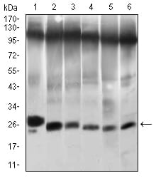
Figure 3: Western blot analysis using TWIST1 mouse mAb against NIH/3T3 (1), JURKAT (2), HELA (3), A549 (4), RAJI (5) and OCM-1 (6) cell lysate.
Immunofluorescence analysis

Figure 4: Immunofluorescence analysis of Hela cells using TWIST1 mouse mAb (green). Blue: DRAQ5 fluorescent DNA dye.
Flow cytometric

Figure 5: Flow cytometric analysis of Hela cells using TWIST1 mouse mAb (green) and negative control (red).
Immunohistochemical analysis

Figure 6: Immunohistochemical analysis of paraffin-embedded cervical cancer tissues using TWIST1 mouse mAb with DAB staining.
Immunohistochemical analysis

Figure 7: Immunohistochemical analysis of paraffin-embedded colon cancer tissues using TWIST1 mouse mAb with DAB staining.
Elisa

Black line: Control Antigen (100 ng); Purple line: Antigen(10ng); Blue line: Antigen (50 ng); Red line: Antigen (100 ng);
For Research Use Only. Not for use in diagnostic procedures.