
TUBA8 Primary Antibody
Item Information
Catalog #
Size
Price
Description
This gene encodes a member of the alpha tubulin protein family. Alpha tubulins are one of two core protein families (alpha and beta tubulins) that heterodimerize and assemble to form microtubules. Mutations in this gene are associated with polymicrogyria and optic nerve hypoplasia. Alternate splicing results in multiple transcript variants.Â
Product Overview
Entrez GenelD
51807
Aliases
TUBAL2
Clone#
2D6
Host / Isotype
Mouse / IgG2b
Species Reactivity
Human, Rat
Immunogen
Purified recombinant fragment of human TUBA8 expressed in E. Coli.
Formulation
Ascitic fluid containing 0.03% sodium azide.
Storage
Store at 4°C short term. Aliquot and store at -20°C long term. Avoid freeze/thaw cycles.
Product Applications
WB (Western Blot)
1/500 - 1/2000
IHC_P(Immunohistochemistry)
1/200 - 1/1000
ICC (Immunocytochemistry)
1/200 - 1/1000
FCM (Flow Cytometry)
1/200 - 1/400
ELISA
1/10000
References
1. Am J Hum Genet. 2009 Nov;85(5):737-44.
2. Am J Hum Genet. 2009 Nov;85(5):628-42.
2. Am J Hum Genet. 2009 Nov;85(5):628-42.
Product Image
Western Blot
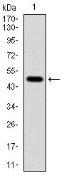
Figure 1: Western blot analysis using TUBA8 mAb against human TUBA8 (AA: 294-449) recombinant protein. (Expected MW is 50 kDa)
Western Blot

Figure 2: Western blot analysis using TUBA8 mouse mAb against rat heart (1) tissue lysate.
Immunohistochemical analysis

Figure 3: Immunohistochemical analysis of paraffin-embedded medulla oblongata tissues using TUBA8 mouse mAb with DAB staining.
Immunohistochemical analysis

Figure 4: Immunohistochemical analysis of paraffin-embedded liver cancer tissues using TUBA8 mouse mAb with DAB staining.
Immunofluorescence analysis

Figure 5: Immunofluorescence analysis of Hela cells using TUBA8 mouse mAb (green). Blue: DRAQ5 fluorescent DNA dye. Red: Actin filaments have been labeled with Alexa Fluor-555 phalloidin.
Flow cytometric

Figure 6: Flow cytometric analysis of NIH/3T3 cells using TUBA8 mouse mAb (green) and negative control (red).
Elisa

Black line: Control Antigen (100 ng); Purple line: Antigen(10ng); Blue line: Antigen (50 ng); Red line: Antigen (100 ng);
For Research Use Only. Not for use in diagnostic procedures.