

TTR Primary Antibody
Item Information
Catalog #
Size
Price
Description
This gene encodes transthyretin, one of the three prealbumins including alpha-1-antitrypsin, transthyretin and orosomucoid. Transthyretin is a carrier protein; it transports thyroid hormones in the plasma and cerebrospinal fluid, and also transports retinol (vitamin A) in the plasma. The protein consists of a tetramer of identical subunits. More than 80 different mutations in this gene have been reported; most mutations are related to amyloid deposition, affecting predominantly peripheral nerve and/or the heart, and a small portion of the gene mutations is non-amyloidogenic. The diseases caused by mutations include amyloidotic polyneuropathy, euthyroid hyperthyroxinaemia, amyloidotic vitreous opacities, cardiomyopathy, oculoleptomeningeal amyloidosis, meningocerebrovascular amyloidosis, carpal tunnel syndrome, etc. [provided by RefSeq, Jan 2009]
Product Overview
Entrez GenelD
7276
Aliases
CTS; CTS1; PALB; TBPA; HEL111; HsT2651
Clone#
2E10C5
Host / Isotype
Mouse / IgG1
Species Reactivity
Human
Immunogen
Purified recombinant fragment of human TTR (AA: 1-147) expressed in E. Coli.
Formulation
Purified antibody from tissue culture in PBS with 0.05% sodium azide
Storage
Store at 4°C short term. Aliquot and store at -20°C long term. Avoid freeze/thaw cycles.
Product Applications
WB (Western Blot)
1/500 - 1/2000
IHC_P(Immunohistochemistry)
1/200 - 1/1000
ICC (Immunocytochemistry)
1/200 - 1/1000
FCM (Flow Cytometry)
1/200 - 1/400
ELISA
1/10000
References
J Biol Chem. 2013 Nov 1;288(44):31752-60.
Clin Exp Rheumatol. 2013 May-Jun;31(3):394-9.
Clin Exp Rheumatol. 2013 May-Jun;31(3):394-9.
Product Image
Elisa
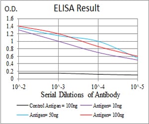
Figure 1: Black line: Control Antigen (100 ng); Purple line: Antigen(10ng); Blue line: Antigen (50 ng); Red line: Antigen (100 ng);
Western Blot
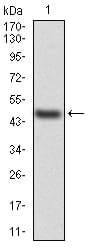
Figure 2:Western blot analysis using TTR mAb against human TTR (AA: 1-147) recombinant protein. (Expected MW is 45.8 kDa)
Western Blot
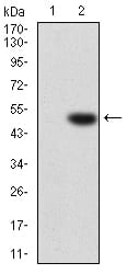
Figure 3:Western blot analysis using TTR mAb against HEK293 (1) and TTR (AA: 1-147)-hIgGFc transfected HEK293 (2) cell lysate.
Immunofluorescence analysis
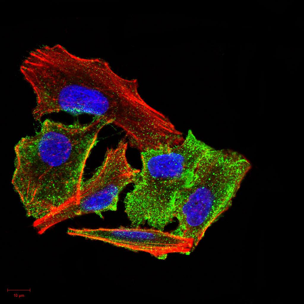
Figure 4:Immunofluorescence analysis of A549 cells using TTR mouse mAb (green). Blue: DRAQ5 fluorescent DNA dye. Red: Actin filaments have been labeled with Alexa Fluor- 555 phalloidin. Secondary antibody from Fisher (Cat#: 35503)
Immunofluorescence analysis
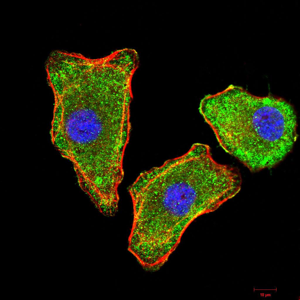
Figure 5:Immunofluorescence analysis of MCF-7 cells using TTR mouse mAb (green). Blue: DRAQ5 fluorescent DNA dye. Red: Actin filaments have been labeled with Alexa Fluor- 555 phalloidin. Secondary antibody from Fisher (Cat#: 35503)
Flow cytometric
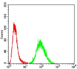
Figure 6:Flow cytometric analysis of A549 cells using TTR mouse mAb (green) and negative control (red).
Immunohistochemical analysis
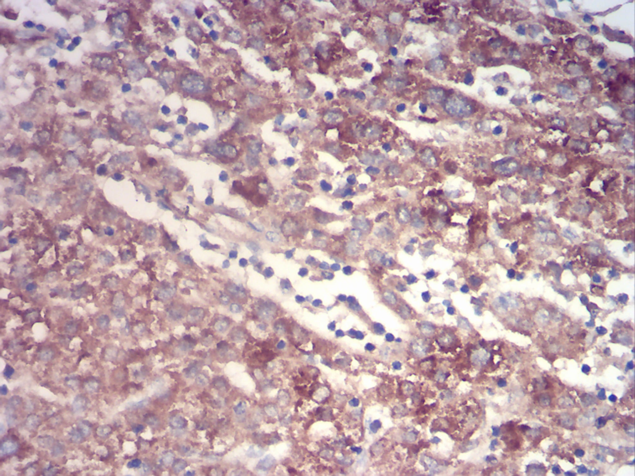
Figure 7:Immunohistochemical analysis of paraffin-embedded liver cancer tissues using TTR mouse mAb with DAB staining.
For Research Use Only. Not for use in diagnostic procedures.