
TTF1 Primary Antibody
Item Information
Catalog #
Size
Price
Description
This gene encodes a transcription termination factor that is localized to the nucleolus and plays a critical role in ribosomal gene transcription. The encoded protein mediates the termination of RNA polymerase I transcription by binding to Sal box terminator elements downstream of pre-rRNA coding regions. Alternatively spliced transcript variants encoding multiple isoforms have been observed for this gene. This gene shares the symbol/alias 'TFF1' with another gene, NK2 homeobox 1, also known as thyroid transcription factor 1, which plays a role in the regulation of thyroid-specific gene expression.
Product Overview
Entrez GenelD
7270
Aliases
TTF-1; TTF-I
Clone#
2F4B12
Host / Isotype
Mouse / IgG1
Species Reactivity
Human
Immunogen
Purified recombinant fragment of human TTF1 (AA: 1-150) expressed in E. Coli.
Formulation
Purified antibody in PBS with 0.05% sodium azide
Storage
Store at 4°C short term. Aliquot and store at -20°C long term. Avoid freeze/thaw cycles.
Product Applications
WB (Western Blot)
1/500 - 1/2000
FCM (Flow Cytometry)
1/200 - 1/400
ELISA
1/10000
References
1.Tumour Biol. 2015 Sep;36(10):8085-92.
2.Chest. 2013 Oct;144(4):1199-206.
2.Chest. 2013 Oct;144(4):1199-206.
Product Image
Elisa
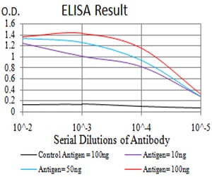
Figure 1: Black line: Control Antigen (100 ng);Purple line: Antigen (10ng); Blue line: Antigen (50 ng); Red line:Antigen (100 ng)
Western Blot
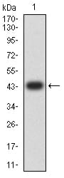
Figure 2:Western blot analysis using TTF1 mAb against human TTF1 (AA: 1-150) recombinant protein. (Expected MW is 43.5 kDa)
Western Blot
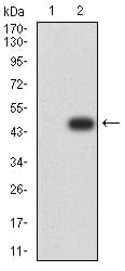
Figure 3:Western blot analysis using TTF1 mAb against HEK293 (1) and TTF1 (AA: 1-150)-hIgGFc transfected HEK293 (2) cell lysate.
Flow cytometric
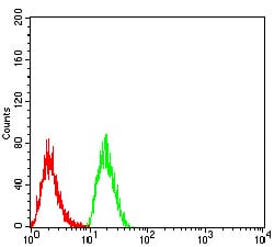
Figure 4:Flow cytometric analysis of Hela cells using TTF1 mouse mAb (green) and negative control (red).
For Research Use Only. Not for use in diagnostic procedures.