
TRIM25 Primary Antibody
Item Information
Catalog #
Size
Price
Description
The protein encoded by this gene is a member of the tripartite motif (TRIM) family. The TRIM motif includes three zinc-binding domains, a RING, a B-box type 1 and a B-box type 2, and a coiled-coil region. The protein localizes to the cytoplasm. The presence of potential DNA-binding and dimerization-transactivation domains suggests that this protein may act as a transcription factor, similar to several other members of the TRIM family. Expression of the gene is upregulated in response to estrogen, and it is thought to mediate estrogen actions in breast cancer as a primary response gene.
Product Overview
Entrez GenelD
7706
Aliases
EFP; Z147; RNF147; ZNF147
Clone#
5B5B12
Host / Isotype
Mouse / IgG2b
Species Reactivity
Human
Immunogen
Purified recombinant fragment of human TRIM25 (AA: 211-360) expressed in E. Coli.
Formulation
Purified antibody in PBS with 0.05% sodium azide
Storage
Store at 4°C short term. Aliquot and store at -20°C long term. Avoid freeze/thaw cycles.
Product Applications
WB (Western Blot)
1/500 - 1/2000
IHC_P(Immunohistochemistry)
1/200 - 1/1000
ICC (Immunocytochemistry)
1/50 - 1/250
FCM (Flow Cytometry)
1/200 - 1/400
ELISA
1/10000
References
1.Science. 2015 Oct 9;350(6257):217-21.
2.Oncogene. 2015 Nov 12;34(46):5729-38.
2.Oncogene. 2015 Nov 12;34(46):5729-38.
Product Image
Elisa
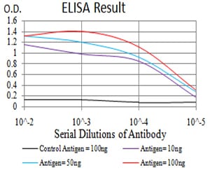
Figure 1: Black line: Control Antigen (100 ng);Purple line: Antigen (10ng); Blue line: Antigen (50 ng); Red line:Antigen (100 ng)
Western Blot
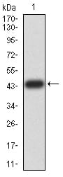
Figure 2:Western blot analysis using TRIM25 mAb against human TRIM25 (AA: 211-360) recombinant protein. (Expected MW is 43.5 kDa)
Western Blot
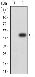
Figure 3:Western blot analysis using TRIM25 mAb against HEK293 (1) and TRIM25 (AA: 211-360)-hIgGFc transfected HEK293 (2) cell lysate.
Western Blot
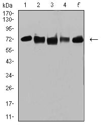
Figure 4:Western blot analysis using TRIM25 mouse mAb against MCF-7 (1), MCF-7 (2), K562 (3), A549 (4), and MOLT4 (5) cell lysate.
Immunofluorescence analysis
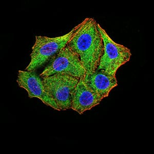
Figure 5:Immunofluorescence analysis of Hela cells using TRIM25 mouse mAb (green). Blue: DRAQ5 fluorescent DNA dye. Red: Actin filaments have been labeled with Alexa Fluor- 555 phalloidin. Secondary antibody from Fisher (Cat#: 35503)
Flow cytometric
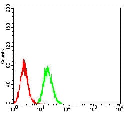
Figure 6:Flow cytometric analysis of Hela cells using TRIM25 mouse mAb (green) and negative control (red).
Immunohistochemical analysis
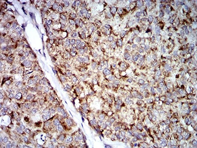
Figure 7:Immunohistochemical analysis of paraffin-embedded bladder cancer tissues using TRIM25 mouse mAb with DAB staining.
Immunohistochemical analysis
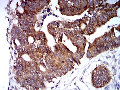
Figure 8:Immunohistochemical analysis of paraffin-embedded rectum cancer tissues using TRIM25 mouse mAb with DAB staining.
For Research Use Only. Not for use in diagnostic procedures.