

Mouse Monoclonal Antibody to TRBC1
Item Information
Catalog #
Size
Price
Description
TRBC1 (T Cell Receptor Beta Constant 1) is a Protein Coding gene. Among its related pathways are Translocation of ZAP-70 to Immunological synapse and Innate Immune System. An important paralog of this gene is TRBC2.
Product Overview
Entrez GenelD
28639
Aliases
TCRB; TCRBC1; BV05S1J2.2
Clone#
5D9F1
Host / Isotype
Mouse / IgG1
Immunogen
Purified recombinant fragment of human TRBC1 (AA: 1-149) expressed in E. Coli.
Formulation
Purified antibody in PBS with 0.05% sodium azide
Storage
Store at 4°C short term. Aliquot and store at -20°C long term. Avoid freeze/thaw cycles.
Product Applications
WB (Western Blot)
1/500 - 1/2000
IHC_P(Immunohistochemistry)
1/200 - 1/1000
ICC (Immunocytochemistry)
1/100 - 1/500
FCM (Flow Cytometry)
1/200 - 1/400
ELISA
1/10000
References
1.Proc Natl Acad Sci U S A. 1985 Aug;82(15):5068-72. 2.Virchows Arch. 2005 Jan;446(1):15-20.
Product Image
Elisa

Figure 1:Black line: Control Antigen (100 ng);Purple line: Antigen (10ng); Blue line: Antigen (50 ng); Red line:Antigen (100 ng)
Western Blot

Figure 2:Western blot analysis using TRBC1 mAb against human TRBC1 (AA: 1-149) recombinant protein. (Expected MW is 42.8 kDa)
Western Blot

Figure 3:Western blot analysis using TRBC1 mAb against HEK293-6e (1) and TRBC1 (AA: 1-149)-hIgGFc transfected HEK293-6e (2) cell lysate.
Western Blot

Figure 4:Western blot analysis using TRBC1 mouse mAb against HUVEC (1), Jurkat (2), Hela (3), HUVE-12 (4), A549 (5), C6 (6), Raji (7), and T47D (8) cell lysate.
Immunofluorescence analysis

Figure 5:Immunofluorescence analysis of Hela cells using TRBC1 mouse mAb (green). Blue: DRAQ5 fluorescent DNA dye. Red: Actin filaments have been labeled with Alexa Fluor- 555 phalloidin. Secondary antibody from Fisher (Cat#: 35503)
Flow cytometric analysis
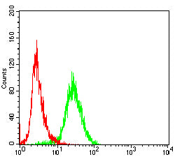
Figure 6:Flow cytometric analysis of Jurkat cells using TRBC1 mouse mAb (green) and negative control (red).
Immunohistochemical analysis

Figure 7:Immunohistochemical analysis of paraffin-embedded testis tissues using TRBC1 mouse mAb with DAB staining.
Immunohistochemical analysis

Figure 8:Immunohistochemical analysis of paraffin-embedded cerebellum tissues using TRBC1 mouse mAb with DAB staining.
For Research Use Only. Not for use in diagnostic procedures.