

TRAF2 Primary Antibody
Item Information
Catalog #
Size
Price
Description
The protein encoded by this gene is a member of the TNF receptor associated factor (TRAF) protein family. TRAF proteins associate with, and mediate the signal transduction from members of the TNF receptor superfamily. This protein directly interacts with TNF receptors, and forms a heterodimeric complex with TRAF1. This protein is required for TNF-alpha-mediated activation of MAPK8/JNK and NF-kappaB. The protein complex formed by this protein and TRAF1 interacts with the inhibitor-of-apoptosis proteins (IAPs), and functions as a mediator of the anti-apoptotic signals from TNF receptors. The interaction of this protein with TRADD, a TNF receptor associated apoptotic signal transducer, ensures the recruitment of IAPs for the direct inhibition of caspase activation. BIRC2/c-IAP1, an apoptosis inhibitor possessing ubiquitin ligase activity, can unbiquitinate and induce the degradation of this protein, and thus potentiate TNF-induced apoptosis. Multiple alternatively spliced transcript variants have been found for this gene, but the biological validity of only one transcript has been determined.
Product Overview
Entrez GenelD
7186
Aliases
TRAP; TRAP3; MGC:45012
Clone#
5C2C3
Host / Isotype
Mouse / IgG1
Species Reactivity
Human
Immunogen
Purified recombinant fragment of human TRAF2 (AA: 39-188) expressed in E. Coli.
Formulation
Purified antibody in PBS with 0.05% sodium azide
Storage
Store at 4°C short term. Aliquot and store at -20°C long term. Avoid freeze/thaw cycles.
Product Applications
WB (Western Blot)
1/500 - 1/2000
IHC_P(Immunohistochemistry)
1/200 - 1/1000
ICC (Immunocytochemistry)
1/200 - 1/1000
FCM (Flow Cytometry)
1/200 - 1/400
ELISA
1/10000
References
1.J Virol. 2014 Apr;88(7):3664-77.
2.Xi Bao Yu Fen Zi Mian Yi Xue Za Zhi. 2011 Nov;27(11):1176-9.
2.Xi Bao Yu Fen Zi Mian Yi Xue Za Zhi. 2011 Nov;27(11):1176-9.
Product Image
Elisa
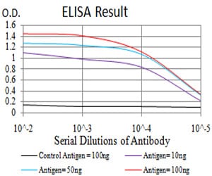
Figure 1: Black line: Control Antigen (100 ng);Purple line: Antigen (10ng); Blue line: Antigen (50 ng); Red line:Antigen (100 ng)
Western Blot
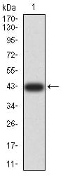
Figure 2:Western blot analysis using TRAF2 mAb against human TRAF2 (AA: 39-188) recombinant protein. (Expected MW is 42.5 kDa)
Western Blot
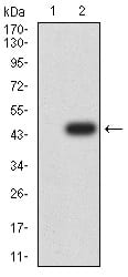
Figure 3:Western blot analysis using TRAF2 mAb against HEK293 (1) and TRAF2 (AA: 39-188)-hIgGFc transfected HEK293 (2) cell lysate.
Immunofluorescence analysis
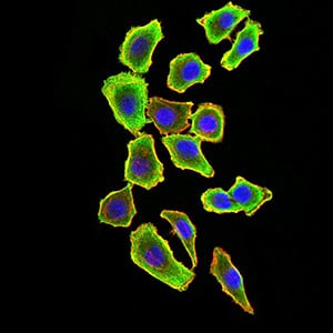
Figure 4:Immunofluorescence analysis of HL-7702 cells using TRAF2 mouse mAb (green). Blue: DRAQ5 fluorescent DNA dye. Red: Actin filaments have been labeled with Alexa Fluor- 555 phalloidin. Secondary antibody from Fisher (Cat#: 35503)
Immunofluorescence analysis
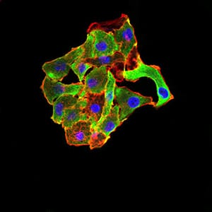
Figure 5:Immunofluorescence analysis of MCF-7 cells using TRAF2 mouse mAb (green). Blue: DRAQ5 fluorescent DNA dye. Red: Actin filaments have been labeled with Alexa Fluor- 555 phalloidin. Secondary antibody from Fisher (Cat#: 35503)
Flow cytometric
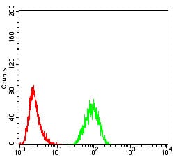
Figure 6:Flow cytometric analysis of Hela cells using TRAF2 mouse mAb (green) and negative control (red).
Flow cytometric
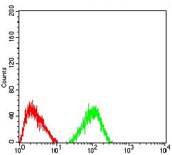
Figure 7:Flow cytometric analysis of HepG2 cells using TRAF2 mouse mAb (green) and negative control (red).
Immunohistochemical analysis
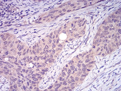
Figure 8:Immunohistochemical analysis of paraffin-embedded cervical cancer tissues using TRAF2 mouse mAb with DAB staining.
For Research Use Only. Not for use in diagnostic procedures.