

TNFSF13B Primary Antibody
Item Information
Catalog #
Size
Price
Description
The protein encoded by this gene is a cytokine that belongs to the tumor necrosis factor (TNF) ligand family. This cytokine is a ligand for receptors TNFRSF13B/TACI, TNFRSF17/BCMA, and TNFRSF13C/BAFFR. This cytokine is expressed in B cell lineage cells, and acts as a potent B cell activator. It has been also shown to play an important role in the proliferation and differentiation of B cells. Alternatively spliced transcript variants encoding distinct isoforms have been identified.
Product Overview
Entrez GenelD
10673
Aliases
DTL; BAFF; BLYS; CD257; TALL1; THANK; ZTNF4; TALL-1; TNLG7A; TNFSF20
Clone#
6C3A2
Host / Isotype
Mouse / IgG1
Species Reactivity
Human
Immunogen
Purified recombinant fragment of human TNFSF13B (AA: 116-278) expressed in E. Coli.
Formulation
Purified antibody in PBS with 0.05% sodium azide
Storage
Store at 4°C short term. Aliquot and store at -20°C long term. Avoid freeze/thaw cycles.
Product Applications
WB (Western Blot)
1/500 - 1/2000
ICC (Immunocytochemistry)
1/100 - 1/500
FCM (Flow Cytometry)
1/200 - 1/400
ELISA
1/10000
References
1.PLoS One. 2015 Nov 23;10(11):e0143393.
2.Biomed Res Int. 2015;2015:792187.
2.Biomed Res Int. 2015;2015:792187.
Product Image
Elisa
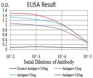
Figure 1: Black line: Control Antigen (100 ng);Purple line: Antigen (10ng); Blue line: Antigen (50 ng); Red line:Antigen (100 ng)
Western Blot
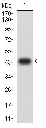
Figure 2:Western blot analysis using TNFSF13B mAb against human TNFSF13B (AA: 116-278) recombinant protein. (Expected MW is 44.1 kDa)
Western Blot
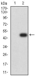
Figure 3:Western blot analysis using TNFSF13B mAb against HEK293 (1) and TNFSF13B (AA: 116-278)-hIgGFc transfected HEK293 (2) cell lysate.
Western Blot
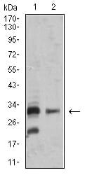
Figure 4:Western blot analysis using TNFSF13B mouse mAb against SK-N-SH (1) and MOLT4 (2) cell lysate.
Immunofluorescence analysis
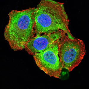
Figure 5:Immunofluorescence analysis of SMMC-7721 cells using TNFSF13B mouse mAb (green). Blue: DRAQ5 fluorescent DNA dye. Red: Actin filaments have been labeled with Alexa Fluor- 555 phalloidin. Secondary antibody from Fisher (Cat#: 35503)
Flow cytometric
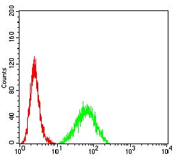
Figure 6:Flow cytometric analysis of HeLa cells using TNFSF13B mouse mAb (green) and negative control (red).
For Research Use Only. Not for use in diagnostic procedures.