

TNFRSF6B Primary Antibody
Item Information
Catalog #
Size
Price
Description
This gene belongs to the tumor necrosis factor receptor superfamily. The encoded protein is postulated to play a regulatory role in suppressing FasL- and LIGHT-mediated cell death. It acts as a decoy receptor that competes with death receptors for ligand binding. Over-expression of this gene has been noted in gastrointestinal tract tumors. Read-through transcription into this gene from the neighboring upstream gene, which encodes regulator of telomere elongation helicase 1 (RTEL1), generates a non-coding transcript.
Product Overview
Entrez GenelD
8771
Aliases
M68; TR6; DCR3; M68E; DJ583P15.1.1
Clone#
3C5H10
Host / Isotype
Mouse / IgG1
Species Reactivity
Human
Immunogen
Purified recombinant fragment of human TNFRSF6B (AA: 30-300) expressed in E. Coli.
Formulation
Purified antibody in PBS with 0.05% sodium azide
Storage
Store at 4°C short term. Aliquot and store at -20°C long term. Avoid freeze/thaw cycles.
Product Applications
WB (Western Blot)
1/500 - 1/2000
FCM (Flow Cytometry)
1/200 - 1/400
ELISA
1/10000
References
1.Chin Med J (Engl). 2016 Nov 5;129(21):2623-2629.2.Sci Rep. 2015 Sep 3;5:12769.
Product Image
Elisa

Figure 1: Black line: Control Antigen (100 ng);Purple line: Antigen (10ng); Blue line: Antigen (50 ng); Red line:Antigen (100 ng)
Western Blot
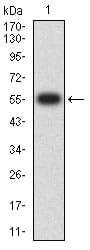
Figure 2:Western blot analysis using TNFRSF6B mAb against human TNFRSF6B (AA: 30-300) recombinant protein. (Expected MW is 55.7 kDa)
Western Blot
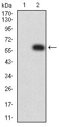
Figure 3:Western blot analysis using TNFRSF6B mAb against HEK293 (1) and TNFRSF6B (AA: 30-300)-hIgGFc transfected HEK293 (2) cell lysate.
Flow cytometric
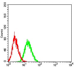
Figure 4:Flow cytometric analysis of HL-60 cells using TNFRSF6B mouse mAb (green) and negative control (red).
For Research Use Only. Not for use in diagnostic procedures.