

TLR9 Primary Antibody
Item Information
Catalog #
Size
Price
Description
The protein encoded by this gene is a member of the Toll-like receptor (TLR) family which plays a fundamental role in pathogen recognition and activation of innate immunity. TLRs are highly conserved from Drosophila to humans and share structural and functional similarities. They recognize pathogen-associated molecular patterns (PAMPs) that are expressed on infectious agents, and mediate the production of cytokines necessary for the development of effective immunity. The various TLRs exhibit different patterns of expression. This gene is preferentially expressed in immune cell rich tissues, such as spleen, lymph node, bone marrow and peripheral blood leukocytes. Studies in mice and human indicate that this receptor mediates cellular response to unmethylated CpG dinucleotides in bacterial DNA to mount an innate immune response.
Product Overview
Entrez GenelD
54106
Aliases
CD289
Clone#
1B12H2
Host / Isotype
Mouse / IgG2a
Species Reactivity
Human
Immunogen
Purified recombinant fragment of human TLR9 (AA: 868-1016) expressed in E. Coli.
Formulation
Purified antibody in PBS with 0.05% sodium azide
Storage
Store at 4°C short term. Aliquot and store at -20°C long term. Avoid freeze/thaw cycles.
Product Applications
WB (Western Blot)
1/500 - 1/2000
IHC_P(Immunohistochemistry)
1/200 - 1/1000
ICC (Immunocytochemistry)
1/200 - 1/1000
FCM (Flow Cytometry)
1/200 - 1/400
ELISA
1/10000
References
1.J Virol. 2015 Nov;89(22):11396-405.
2.Immunogenetics. 2014 Dec;66(12):675-81.
2.Immunogenetics. 2014 Dec;66(12):675-81.
Product Image
Elisa
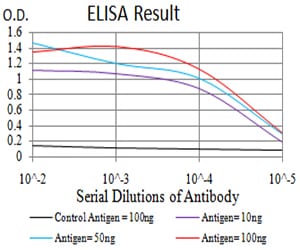
Figure 1: Black line: Control Antigen (100 ng);Purple line: Antigen (10ng); Blue line: Antigen (50 ng); Red line:Antigen (100 ng)
Western Blot

Figure 2:Western blot analysis using TLR9 mAb against human TLR9 (AA: 868-1016) recombinant protein. (Expected MW is 43.4 kDa)
Western Blot

Figure 3:Western blot analysis using TLR9 mAb against HEK293 (1) and TLR9 (AA: 868-1016)-hIgGFc transfected HEK293 (2) cell lysate.
Immunofluorescence analysis

Figure 4:Immunofluorescence analysis of SK-OV-3 cells using TLR9 mouse mAb (green). Blue: DRAQ5 fluorescent DNA dye. Red: Actin filaments have been labeled with Alexa Fluor- 555 phalloidin. Secondary antibody from Fisher (Cat#: 35503)
Flow cytometric

Figure 5:Flow cytometric analysis of A549 cells using TLR9 mouse mAb (green) and negative control (red).
Immunohistochemical analysis

Figure 6:Immunohistochemical analysis of paraffin-embedded ovarian cancer tissues using TLR9 mouse mAb with DAB staining.
Immunohistochemical analysis

Figure 7:Immunohistochemical analysis of paraffin-embedded endometrial cancer tissues using TLR9 mouse mAb with DAB staining.
For Research Use Only. Not for use in diagnostic procedures.