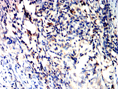
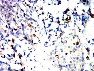
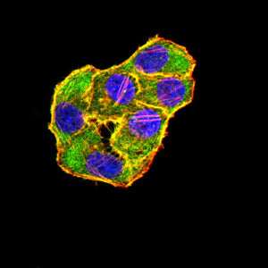
Mouse Monoclonal Antibody to TLR10
The protein encoded by this gene is a member of the Toll-like receptor (TLR) family which plays a fundamental role in pathogen recognition and activation of innate immunity. TLRs are highly conserved from Drosophila to humans and share structural and functional similarities. They recognize pathogen-associated molecular patterns (PAMPs) that are expressed on infectious agents, and mediate the production of cytokines necessary for the development of effective immunity. The various TLRs exhibit different patterns of expression. This gene is most highly expressed in lymphoid tissues such as spleen, lymph node, thymus, and tonsil. Multiple alternatively spliced transcript variants which encode different protein isoforms have been found for this gene. [provided by RefSeq, Aug 2010]
![Figure 1:Black line: Control Antigen (100 ng);Purple line: Antigen (10ng); Blue line: Antigen (50 ng); Red line:Antigen (100 ng)](https://cdn.shopify.com/s/files/1/0604/2661/7019/products/9165ae30d1ee995562d360e4f466951349ac420d860d49cedf90b763de3cd3a4.jpg?v=1666705737)
![Figure 2:Western blot analysis using TLR10 mAb against human TLR10 (AA: extra(20-219)) recombinant protein. (Expected MW is 26kDa)](https://cdn.shopify.com/s/files/1/0604/2661/7019/products/98265879665bcd0b9cad543697ed11767c9a7dc32c79f736deb2fec1d7ef059b.jpg?v=1666705737)
![Figure 3:Western blot analysis using TLR10 mAb against HEK293-6e (1) and TLR10 (AA: extra(20-219))-hIgGFc transfected HEK293-6e (2) cell lysate.](https://cdn.shopify.com/s/files/1/0604/2661/7019/products/28f326418c6a5d5e83523f682473c6bde2731263a4e71a040aa805b369bb5ad8.jpg?v=1666705737)
![Figure 4:Flow cytometric analysis of Jurkat cells using TLR10 mouse mAb (green) and negative control (red).](https://cdn.shopify.com/s/files/1/0604/2661/7019/products/adf4fad88b6788046f4e058a2671d183b5d7192eb952329a467f4123bc01a85f.jpg?v=1666705737)
![Figure 5:Immunohistochemical analysis of paraffin-embedded cervical cancer tissues using TLR10 mouse mAb with DAB staining.](https://cdn.shopify.com/s/files/1/0604/2661/7019/products/9351dde74f817bce8559f581fefe80d11b12de46c2ff8bde3aa1387ae196bc6d.jpg?v=1666705737)
![Figure 6:Immunohistochemical analysis of paraffin-embedded esophageal cancer tissues using TLR10 mouse mAb with DAB staining.](https://cdn.shopify.com/s/files/1/0604/2661/7019/products/1ef5308486285c82779590bf9505e07716e9848132212c8fc3482e45d5393f11.jpg?v=1666705737)
![Figure 7:Immunofluorescence analysis of Hela cells using TLR10 mouse mAb (green). Blue: DRAQ5 fluorescent DNA dye. Red: Actin filaments have been labeled with Alexa Fluor- 555 phalloidin. Secondary antibody from Fisher (Cat#: 35503)](https://cdn.shopify.com/s/files/1/0604/2661/7019/products/ae29ca18a3229f0a05254f30bf3004131707a598e764d22004ca53c94fec993a.jpg?v=1666705737)