
TLL1 Primary Antibody
Item Information
Catalog #
Size
Price
Description
This gene encodes an astacin-like, zinc-dependent, metalloprotease that belongs to the peptidase M12A family. This protease processes procollagen C-propeptides, such as chordin, pro-biglycan and pro-lysyl oxidase. Studies in mice suggest that this gene plays multiple roles in the development of mammalian heart, and is essential for the formation of the interventricular septum. Allelic variants of this gene are associated with atrial septal defect type 6. Alternatively spliced transcript variants encoding different isoforms have been found for this gene.
Product Overview
Entrez GenelD
7092
Aliases
TLL; ASD6
Clone#
4H8C1
Host / Isotype
Mouse / IgG1
Species Reactivity
Human
Immunogen
Purified recombinant fragment of human TLL1 (AA: 870-1013) expressed in E. Coli.
Formulation
Purified antibody in PBS with 0.05% sodium azide
Storage
Store at 4°C short term. Aliquot and store at -20°C long term. Avoid freeze/thaw cycles.
Product Applications
WB (Western Blot)
1/500 - 1/2000
FCM (Flow Cytometry)
1/200 - 1/400
ELISA
1/10000
References
1.Zhonghua Xin Xue Guan Bing Za Zhi. 2012 May;40(5):402-5.
2.Biochem Biophys Res Commun. 2009 Nov 13;389(2):338-42.
2.Biochem Biophys Res Commun. 2009 Nov 13;389(2):338-42.
Product Image
Elisa

Figure 1: Black line: Control Antigen (100 ng);Purple line: Antigen (10ng); Blue line: Antigen (50 ng); Red line:Antigen (100 ng)
Western Blot

Figure 2:Western blot analysis using TLL1 mAb against human TLL1 (AA: 870-1013) recombinant protein. (Expected MW is 42.2 kDa)
Western Blot

Figure 3:Western blot analysis using TLL1 mAb against HEK293 (1) and TLL1 (AA: 870-1013)-hIgGFc transfected HEK293 (2) cell lysate.
Flow cytometric
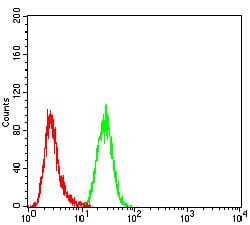
Figure 4:Flow cytometric analysis of Hela cells using TLL1 mouse mAb (green) and negative control (red).
For Research Use Only. Not for use in diagnostic procedures.