

TIP60 Primary Antibody
Item Information
Catalog #
Size
Price
Description
The protein encoded by this gene belongs to the MYST family of histone acetyl transferases (HATs) and was originally isolated as an HIV-1 TAT-interactive protein. HATs play important roles in regulating chromatin remodeling, transcription and other nuclear processes by acetylating histone and nonhistone proteins. This protein is a histone acetylase that has a role in DNA repair and apoptosis and is thought to play an important role in signal transduction. Alternative splicing of this gene results in multiple transcript variants.
Product Overview
Entrez GenelD
10524
Aliases
TIP; ESA1; PLIP; KAT5; cPLA2; HTATIP; ZC2HC5; HTATIP1
Clone#
2A11H7
Host / Isotype
Mouse / IgG1
Species Reactivity
Human
Immunogen
Purified recombinant fragment of human TIP60 (AA: 18-208) expressed in E. Coli.
Formulation
Purified antibody from tissue culture in PBS with 0.05% sodium azide
Storage
Store at 4°C short term. Aliquot and store at -20°C long term. Avoid freeze/thaw cycles.
Product Applications
WB (Western Blot)
1/500 - 1/2000
FCM (Flow Cytometry)
1/200 - 1/400
ELISA
1/10000
References
Anticancer Res. 2011 Jan;31(1):77-9.
J Invest Dermatol. 2012 Nov;132(11):2632-41.
J Invest Dermatol. 2012 Nov;132(11):2632-41.
Product Image
Elisa
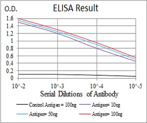
Figure 1: Black line: Control Antigen (100 ng); Purple line: Antigen(10ng); Blue line: Antigen (50 ng); Red line: Antigen (100 ng);
Western Blot
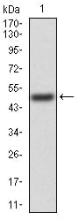
Figure 2:Western blot analysis using TIP60 mAb against human TIP60 (AA: 18-208) recombinant protein. (Expected MW is 47.9 kDa)
Western Blot
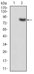
Figure 3:Western blot analysis using TIP60 mAb against HEK293 (1) and TIP60 (AA: 18-208)-hIgGFc transfected HEK293 (2) cell lysate.
Western Blot

Figure 4:Western blot analysis using TIP60 mouse mAb against Hela (1) cell lysate.
Flow cytometric
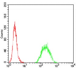
Figure 5:Flow cytometric analysis of Hela cells using TIP60 mouse mAb (green) and negative control (red).
For Research Use Only. Not for use in diagnostic procedures.