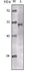
TIP60 Primary Antibody
HTATIP (HIV-1 Tat interacting protein TIP60, about 60kDa) belongs to the MYST family of histone acetyl transferases (HATs) and was originally isolated as an HIV-1 TAT-interactive protein. HATs play important roles in regulating chromatin remodeling, transcription and other nuclear processes by acetylating histone and nonhistone proteins. The nucleosome, made up of four core histone proteins (H2A, H2B, H3 and H4), is the primary building block of chromatin. In addition to the growing number of post-translational histone modifications regulating chromatin structure, cells can also exchange canonical histones with variant histones that can directly or indirectly modulate chromatin structure. There are five major variants of histone H2A: canonical H2A (most abundant), H2A.X, MacroH2A, H2ABbd and H2A.Z. Histone H2A.Z, the most conserved variant across species, functions as both a positive and negative regulator of transcription and is important for chromosome stability. Several homologous protein complexes, such as SWR-C, TIP60 and SRCAP (mammals), have been shown to catalyze the ATP-dependent exchange of H2A.Z for H2A in the nucleosome. This protein is a histone acetylase that has a role in DNA repair and apoptosis and is thought to play an important role in signal transduction.
2. Jin, J. et al. Trends Biochem. Sci. 2005 30, 680-687.
3. Raisner, R.M. and Madhani, H.D. Curr. Opin. Genet. 2006 Dev. 16, 119-124.