
TIE1 Primary Antibody
Item Information
Catalog #
Size
Price
Description
This gene encodes a member of the tyrosine protein kinase family. The encoded protein plays a critical role in angiogenesis and blood vessel stability by inhibiting angiopoietin 1 signaling through the endothelial receptor tyrosine kinase Tie2. Ectodomain cleavage of the encoded protein relieves inhibition of Tie2 and is mediated by multiple factors including vascular endothelial growth factor. Alternatively spliced transcript variants encoding multiple isoforms have been observed for this gene.
Product Overview
Entrez GenelD
7075
Aliases
TIE; JTK14
Clone#
8D12D2
Host / Isotype
Mouse / IgG1
Species Reactivity
Human
Immunogen
Purified recombinant fragment of human TIE1 (AA: 385-607) expressed in E. Coli.
Formulation
Purified antibody in PBS with 0.05% sodium azide.
Storage
Store at 4°C short term. Aliquot and store at -20°C long term. Avoid freeze/thaw cycles.
Product Applications
WB (Western Blot)
1/500 - 1/2000
IHC_P(Immunohistochemistry)
1/200 - 1/1000
ELISA
1/10000
References
1. Int J Oncol. 2007 Oct;31(4):893-7.
2. Cancer. 2002 Mar 1;94(5):1517-21.
2. Cancer. 2002 Mar 1;94(5):1517-21.
Product Image
Western Blot

Figure 1: Western blot analysis using TIE1 mAb against human TIE1 (AA: 385-607) recombinant protein. (Expected MW is 50.6 kDa)
Western Blot
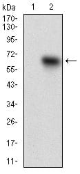
Figure 2: Western blot analysis using TIE1 mAb against HEK293 (1) and TIE1 (AA: 385-607)-hIgGFc transfected HEK293 (2) cell lysate.
Western Blot

Figure 3: Western blot analysis using TIE1 mouse mAb against HepG2 cell lysate.
Immunohistochemical analysis

Figure 4: Immunohistochemical analysis of paraffin-embedded ovarian cancer tissues using TIE1 mouse mAb with DAB staining.
Immunohistochemical analysis

Figure 5: Immunohistochemical analysis of paraffin-embedded kidney tissues using TIE1 mouse mAb with DAB staining.
Elisa
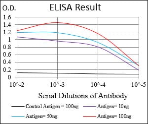
Black line: Control Antigen (100 ng); Purple line: Antigen(10ng); Blue line: Antigen (50 ng); Red line: Antigen (100 ng);
For Research Use Only. Not for use in diagnostic procedures.