
THAP1 Primary Antibody
Item Information
Catalog #
Size
Price
Description
The protein encoded by this gene contains a THAP domain, a conserved DNA-binding domain. This protein colocalizes with the apoptosis response protein PAWR/PAR-4 in promyelocytic leukemia (PML) nuclear bodies, and functions as a proapoptotic factor that links PAWR to PML nuclear bodies. Alternatively spliced transcript variants encoding distinct isoforms have been observed.
Product Overview
Entrez GenelD
55145
Aliases
DYT6
Clone#
2F1B4
Host / Isotype
Mouse / IgG1
Species Reactivity
Human
Immunogen
Purified recombinant fragment of human THAP1 (AA: 1-213) expressed in E. Coli.
Formulation
Purified antibody in PBS with 0.05% sodium azide
Storage
Store at 4°C short term. Aliquot and store at -20°C long term. Avoid freeze/thaw cycles.
Product Applications
WB (Western Blot)
1/500 - 1/2000
FCM (Flow Cytometry)
1/200 - 1/400
ELISA
1/10000
References
1.Mov Disord. 2014 Feb;29(2):278-80.
2.Hum Mutat. 2011 Nov;32(11):1213-24.
2.Hum Mutat. 2011 Nov;32(11):1213-24.
Product Image
Elisa
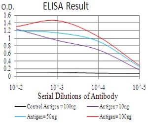
Figure 1: Black line: Control Antigen (100 ng); Purple line: Antigen(10ng); Blue line: Antigen (50 ng); Red line: Antigen (100 ng);
Western Blot

Figure 2:Western blot analysis using THAP1 mAb against human THAP1 (AA: 1-213) recombinant protein. (Expected MW is 50.3 kDa)
Western Blot
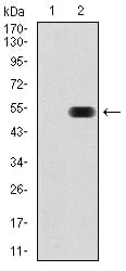
Figure 3:Western blot analysis using THAP1 mAb against HEK293 (1) and THAP1 (AA: 1-213)-hIgGFc transfected HEK293 (2) cell lysate.
Flow cytometric

Figure 4:Flow cytometric analysis of A549 cells using THAP1 mouse mAb (green) and negative control (red).
For Research Use Only. Not for use in diagnostic procedures.