
TFAP2A Primary Antibody
Item Information
Catalog #
Size
Price
Description
The protein encoded by this gene is a transcription factor that binds the consensus sequence 5'-GCCNNNGGC-3'. The encoded protein functions as either a homodimer or as a heterodimer with similar family members. This protein activates the transcription of some genes while inhibiting the transcription of others. Defects in this gene are a cause of branchiooculofacial syndrome (BOFS). Three transcript variants encoding different isoforms have been found for this gene.
Product Overview
Entrez GenelD
7020
Aliases
AP-2; BOFS; AP2TF; TFAP2; AP-2alpha
Clone#
1A10C5
Host / Isotype
Mouse / IgG1
Species Reactivity
Human
Immunogen
Purified recombinant fragment of human TFAP2A (AA: 1-100) expressed in E. Coli.
Formulation
Purified antibody in PBS with 0.05% sodium azide
Storage
Store at 4°C short term. Aliquot and store at -20°C long term. Avoid freeze/thaw cycles.
Product Applications
WB (Western Blot)
1/500 - 1/2000
ICC (Immunocytochemistry)
1/200 - 1/1000
FCM (Flow Cytometry)
1/200 - 1/400
ELISA
1/10000
References
Mol Hum Reprod. 2011 Nov;17(11):702-9.
Breast Cancer Res. 2011 Mar 4;13(2):R23.
Breast Cancer Res. 2011 Mar 4;13(2):R23.
Product Image
Elisa
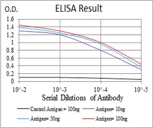
Figure 1: Black line: Control Antigen (100 ng); Purple line: Antigen(10ng); Blue line: Antigen (50 ng); Red line: Antigen (100 ng);
Western Blot
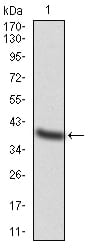
Figure 2:Western blot analysis using TFAP2A mAb against human TFAP2A (AA: 1-100) recombinant protein. (Expected MW is 37 kDa)
Western Blot

Figure 3:Western blot analysis using TFAP2A mAb against HEK293 (1) and TFAP2A (AA: 1-100)-hIgGFc transfected HEK293 (2) cell lysate.
Immunofluorescence analysis

Figure 4:Immunofluorescence analysis of Hela cells using TFAP2A mouse mAb (green). Blue: DRAQ5 fluorescent DNA dye. Red: Actin filaments have been labeled with Alexa Fluor- 555 phalloidin. Secondary antibody from Fisher (Cat#: 35503)
Flow cytometric

Figure 5:Flow cytometric analysis of Hela cells using TFAP2A mouse mAb (green) and negative control (red).
For Research Use Only. Not for use in diagnostic procedures.