

TCL1A Primary Antibody
Item Information
Catalog #
Size
Price
Description
Overexpression of the TCL1 gene in humans has been implicated in the development of mature T cell leukemia, in which chromosomal rearrangements bring the TCL1 gene in close proximity to the T-cell antigen receptor (TCR)-alpha (MIM 186880) or TCR-beta (MIM 186930) regulatory elements (summarized by Virgilio et al., 1998 [PubMed 9520462]). In normal T cells TCL1 is expressed in CD4-/CD8- cells, but not in cells at later stages of differentiation. TCL1 functions as a coactivator of the cell survival kinase AKT (MIM 164730) (Laine et al., 2000 [PubMed 10983986]).
Product Overview
Entrez GenelD
8115
Aliases
TCL1
Clone#
2E3A5
Host / Isotype
Mouse / IgG1
Species Reactivity
Human
Immunogen
Purified recombinant fragment of human TCL1A (AA: 10-104) expressed in E. Coli.
Formulation
Purified antibody from tissue culture in PBS with 0.05% sodium azide
Storage
Store at 4°C short term. Aliquot and store at -20°C long term. Avoid freeze/thaw cycles.
Product Applications
WB (Western Blot)
1/500 - 1/2000
FCM (Flow Cytometry)
1/200 - 1/400
ELISA
1/10000
References
1. Blood. 2012 Aug 23;120(8):1613-23.
2. Histopathology. 2010 Jul;57(1):152-7.
2. Histopathology. 2010 Jul;57(1):152-7.
Product Image
Western Blot
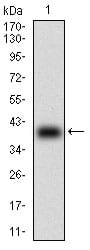
Figure 1: Western blot analysis using TCL1A mAb against human TCL1A (AA: 10-104) recombinant protein. (Expected MW is 37.3 kDa)
Western Blot
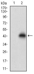
Figure 2: Western blot analysis using TCL1A mAb against HEK293 (1) and TCL1A (AA: 10-104)-hIgGFc transfected HEK293 (2) cell lysate.
Flow cytometric
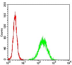
Figure 3: Flow cytometric analysis of Hela cells using TCL1A mouse mAb (green) and negative control (red).
Elisa
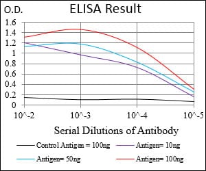
Black line: Control Antigen (100 ng); Purple line: Antigen(10ng); Blue line: Antigen (50 ng); Red line: Antigen (100 ng);
For Research Use Only. Not for use in diagnostic procedures.