
TCF7 Primary Antibody
Item Information
Catalog #
Size
Price
Description
This gene encodes a member of the T-cell factor/lymphoid enhancer-binding factor family of high mobility group (HMG) box transcriptional activators. This gene is expressed predominantly in T-cells and plays a critical role in natural killer cell and innate lymphoid cell development. The encoded protein forms a complex with beta-catenin and activates transcription through a Wnt/beta-catenin signaling pathway. Mice with a knockout of this gene are viable and fertile, but display a block in T-lymphocyte differentiation. Alternative splicing results in multiple transcript variants. Naturally-occurring isoforms lacking the N-terminal beta-catenin interaction domain may act as dominant negative regulators of Wnt signaling.
Product Overview
Entrez GenelD
6932
Aliases
TCF-1
Clone#
1B4E9
Host / Isotype
Mouse / Mouse IgG1
Species Reactivity
Human
Immunogen
Purified recombinant fragment of human TCF7 (AA: 168-358) expressed in E. Coli.
Formulation
Purified antibody in PBS with 0.05% sodium azide
Storage
Store at 4°C short term. Aliquot and store at -20°C long term. Avoid freeze/thaw cycles.
Product Applications
WB (Western Blot)
1/500 - 1/2000
IHC_P(Immunohistochemistry)
1/200 - 1/1000
ICC (Immunocytochemistry)
N/A
FCM (Flow Cytometry)
1/200 - 1/400
ELISA
1/10000
References
1.J Pak Med Assoc. 2020 Oct;70(10):1774-1778.
2.Med Sci Monit. 2019 May 28;25:3957-3963.
2.Med Sci Monit. 2019 May 28;25:3957-3963.
Product Image
Elisa

Figure 1:Black line: Control Antigen (100 ng);Purple line: Antigen (10ng); Blue line: Antigen (50 ng); Red line:Antigen (100 ng)
Western Blot
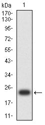
Figure 2:Western blot analysis using TCF7 mAb against human TCF7 (AA: 168-358) recombinant protein. (Expected MW is 24.5 kDa)
Western Blot

Figure 3:Western blot analysis using TCF7 mAb against HEK293-6e (1) and TCF7 (AA:168-358)-hIgGFc transfected HEK293-6e (2) cell lysate.
Immunofluorescence analysis
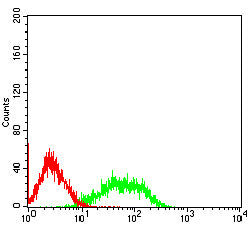
Figure 4:Flow cytometric analysis of Jurkat cells using TCF7 mouse mAb (green) and negative control (red).
Immunohistochemical analysis

Figure 5:Immunohistochemical analysis of paraffin-embedded ovarian cancer tissues using TCF7 mouse mAb with DAB staining.
Immunohistochemical analysis

Figure 6:Immunohistochemical analysis of paraffin-embedded rectum cancer tissues using TCF7 mouse mAb with DAB staining.
For Research Use Only. Not for use in diagnostic procedures.