

TCF4 Primary Antibody
Item Information
Catalog #
Size
Price
Description
This gene encodes transcription factor 4, a basic helix-loop-helix transcription factor. The encoded protein recognizes an Ephrussi-box ('E-box') binding site ('CANNTG') - a motif first identified in immunoglobulin enhancers. This gene is broadly expressed, and may play an important role in nervous system development. Defects in this gene are a cause of Pitt-Hopkins syndrome. In addition, an intronic CTG repeat normally numbering 10-37 repeat units can expand to >50 repeat units and cause Fuchs endothelial corneal dystrophy. Multiple alternatively spliced transcript variants that encode different proteins have been described.
Product Overview
Entrez GenelD
6925
Aliases
E2-2; ITF2; PTHS; SEF2; FECD3; ITF-2; SEF-2; TCF-4
Clone#
4D4C4
Host / Isotype
Mouse / IgG1
Species Reactivity
Human
Immunogen
Purified recombinant fragment of human TCF4 (AA: 518-667) expressed in E. Coli.
Formulation
Purified antibody in PBS with 0.05% sodium azide
Storage
Store at 4°C short term. Aliquot and store at -20°C long term. Avoid freeze/thaw cycles.
Product Applications
WB (Western Blot)
1/500 - 1/2000
FCM (Flow Cytometry)
1/200 - 1/400
ELISA
1/10000
References
1.J Psychiatr Res. 2015 Oct;69:95-101.
2.Haematologica. 2014 Dec;99(12):e257-9.
2.Haematologica. 2014 Dec;99(12):e257-9.
Product Image
Elisa
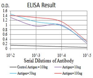
Figure 1: Black line: Control Antigen (100 ng);Purple line: Antigen (10ng); Blue line: Antigen (50 ng); Red line:Antigen (100 ng)
Western Blot

Figure 2:Western blot analysis using TCF4 mAb against human TCF4 (AA: 518-667) recombinant protein. (Expected MW is 43 kDa)
Western Blot
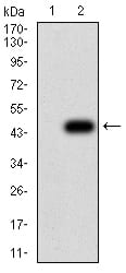
Figure 3:Western blot analysis using TCF4 mAb against HEK293 (1) and TCF4 (AA: 518-667)-hIgGFc transfected HEK293 (2) cell lysate.
Flow cytometric

Figure 4:Flow cytometric analysis of K562 cells using TCF4 mouse mAb (green) and negative control (red).
For Research Use Only. Not for use in diagnostic procedures.