

TBC1D4 Primary Antibody
Item Information
Catalog #
Size
Price
Description
This gene is a member of the Tre-2/BUB2/CDC16 domain family. The protein encoded by this gene is a Rab-GTPase-activating protein, and contains two phopshotyrosine-binding domains (PTB1 and PTB2), a calmodulin-binding domain (CBD), a Rab-GTPase domain, and multiple AKT phosphomotifs. This protein is thought to play an important role in glucose homeostasis by regulating the insulin-dependent trafficking of the glucose transporter 4 (GLUT4), important for removing glucose from the bloodstream into skeletal muscle and fat tissues. Reduced expression of this gene results in an increase in GLUT4 levels at the plasma membrane, suggesting that this protein is important in intracellular retention of GLUT4 under basal conditions. When exposed to insulin, this protein is phosphorylated, dissociates from GLUT4 vesicles, resulting in increased GLUT4 at the cell surface, and enhanced glucose transport. Phosphorylation of this protein by AKT is required for proper translocation of GLUT4 to the cell surface. Individuals homozygous for a mutation in this gene are at higher risk for type 2 diabetes and have higher levels of circulating glucose and insulin levels after glucose ingestion. Alternative splicing results in multiple transcript variants encoding different isoforms.
Product Overview
Entrez GenelD
9882
Aliases
AS160; NIDDM5
Clone#
8A11A2
Host / Isotype
Mouse / IgG1
Species Reactivity
Human
Immunogen
Purified recombinant fragment of human TBC1D4 (AA: 574-712) expressed in E. Coli.
Formulation
Purified antibody in PBS with 0.05% sodium azide
Storage
Store at 4°C short term. Aliquot and store at -20°C long term. Avoid freeze/thaw cycles.
Product Applications
WB (Western Blot)
1/500 - 1/2000
FCM (Flow Cytometry)
1/200 - 1/400
ELISA
1/10000
References
1.Am J Physiol Endocrinol Metab. 2012 Jan 15;302(2):E190-200.
2.Cancer Biol Ther. 2010 Aug 15;10(4):362-7.
2.Cancer Biol Ther. 2010 Aug 15;10(4):362-7.
Product Image
Elisa
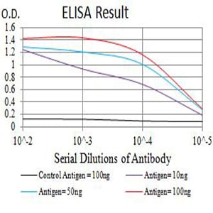
Figure 1: Black line: Control Antigen (100 ng);Purple line: Antigen (10ng); Blue line: Antigen (50 ng); Red line:Antigen (100 ng)
Western Blot

Figure 2:Western blot analysis using TBC1D4 mAb against human TBC1D4 (AA: 574-712) recombinant protein. (Expected MW is 41.2 kDa)
Western Blot

Figure 3:Western blot analysis using TBC1D4 mAb against HEK293 (1) and TBC1D4 (AA: 574-712)-hIgGFc transfected HEK293 (2) cell lysate.
Flow cytometric

Figure 4:Flow cytometric analysis of Hela cells using TBC1D4 mouse mAb (green) and negative control (red).
For Research Use Only. Not for use in diagnostic procedures.