

SYN1 Primary Antibody
Item Information
Catalog #
Size
Price
Description
This gene is a member of the synapsin gene family. Synapsins encode neuronal phosphoproteins which associate with the cytoplasmic surface of synaptic vesicles. Family members are characterized by common protein domains, and they are implicated in synaptogenesis and the modulation of neurotransmitter release, suggesting a potential role in several neuropsychiatric diseases. This member of the synapsin family plays a role in regulation of axonogenesis and synaptogenesis. The protein encoded serves as a substrate for several different protein kinases and phosphorylation may function in the regulation of this protein in the nerve terminal. Mutations in this gene may be associated with X-linked disorders with primary neuronal degeneration such as Rett syndrome. Alternatively spliced transcript variants encoding different isoforms have been identified.
Product Overview
Entrez GenelD
6853
Aliases
SYNI; SYN1a; SYN1b
Clone#
7B1D9
Host / Isotype
Mouse / IgG1
Species Reactivity
Human, Mouse, Rat
Immunogen
Purified recombinant fragment of human SYN1 (AA: 362-511) expressed in E. Coli.
Formulation
Purified antibody in PBS with 0.05% sodium azide
Storage
Store at 4°C short term. Aliquot and store at -20°C long term. Avoid freeze/thaw cycles.
Product Applications
WB (Western Blot)
1/500 - 1/2000
ICC (Immunocytochemistry)
1/200 - 1/1000
ELISA
1/10000
References
1.Synapse. 2012 Nov;66(11):979-83.
2.J Neurosci Res. 2009 Aug 1;87(10):2255-63.
2.J Neurosci Res. 2009 Aug 1;87(10):2255-63.
Product Image
Elisa
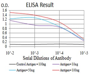
Figure 1: Black line: Control Antigen (100 ng);Purple line: Antigen (10ng); Blue line: Antigen (50 ng); Red line:Antigen (100 ng)
Western Blot
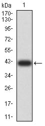
Figure 2:Western blot analysis using SYN1 mAb against human SYN1 (AA: 362-511) recombinant protein. (Expected MW is 41.7 kDa)
Western Blot
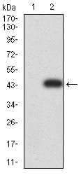
Figure 3:Western blot analysis using SYN1 mAb against HEK293 (1) and SYN1 (AA: 362-511)-hIgGFc transfected HEK293 (2) cell lysate.
Western Blot
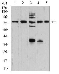
Figure 4:Western blot analysis using SYN1 mouse mAb against NIH/3T3 (1), U251 (2), C6 (3), A549 (4), and MCF-7 (5) cell lysate.
Immunofluorescence analysis
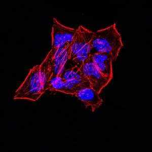
Figure 5:Immunofluorescence analysis of Hela cells. Blue: DRAQ5 fluorescent DNA dye. Red: Actin filaments have been labeled with Alexa Fluor- 555 phalloidin. Secondary antibody from Fisher (Cat#: 35503)
Immunofluorescence analysis
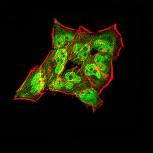
Figure 6:Immunofluorescence analysis of Hela cells using SYN1 mouse mAb (green). Blue: DRAQ5 fluorescent DNA dye. Red: Actin filaments have been labeled with Alexa Fluor- 555 phalloidin. Secondary antibody from Fisher (Cat#: 35503)
Immunofluorescence analysis

Figure 7:Immunofluorescence analysis of HepG2 cells. Blue: DRAQ5 fluorescent DNA dye. Red: Actin filaments have been labeled with Alexa Fluor- 555 phalloidin. Secondary antibody from Fisher (Cat#: 35503)
Immunofluorescence analysis

Figure 8:Immunofluorescence analysis of HepG2 cells using SYN1 mouse mAb (green). Blue: DRAQ5 fluorescent DNA dye. Red: Actin filaments have been labeled with Alexa Fluor- 555 phalloidin. Secondary antibody from Fisher (Cat#: 35503)
For Research Use Only. Not for use in diagnostic procedures.