

SYK Primary Antibody
Item Information
Catalog #
Size
Price
Description
This gene encodes a member of the family of non-receptor type Tyr protein kinases. This protein is widely expressed in hematopoietic cells and is involved in coupling activated immunoreceptors to downstream signaling events that mediate diverse cellular responses, including proliferation, differentiation, and phagocytosis. It is thought to be a modulator of epithelial cell growth and a potential tumour suppressor in human breast carcinomas. Alternatively spliced transcript variants encoding different isoforms have been found for this gene.
Product Overview
Entrez GenelD
6850
Aliases
p72-Syk
Clone#
2G11E6
Host / Isotype
Mouse / IgG1
Species Reactivity
Human
Immunogen
Purified recombinant fragment of human SYK (AA: 217-356) expressed in E. Coli.
Formulation
Purified antibody in PBS with 0.05% sodium azide
Storage
Store at 4°C short term. Aliquot and store at -20°C long term. Avoid freeze/thaw cycles.
Product Applications
WB (Western Blot)
1/500 - 1/2000
ICC (Immunocytochemistry)
1/50 - 1/250
FCM (Flow Cytometry)
1/200 - 1/400
ELISA
1/10000
References
1.Clin Cancer Res. 2015 Jun 1;21(11):2538-45.
2.PLoS One. 2014 Feb 11;9(2):e87610.
2.PLoS One. 2014 Feb 11;9(2):e87610.
Product Image
Elisa
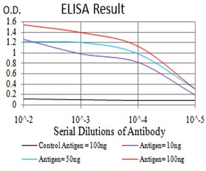
Figure 1: Black line: Control Antigen (100 ng);Purple line: Antigen (10ng); Blue line: Antigen (50 ng); Red line:Antigen (100 ng)
Western Blot
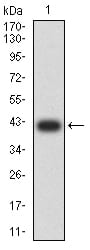
Figure 2:Western blot analysis using SYK mAb against human SYK (AA: 217-356) recombinant protein. (Expected MW is 41.4 kDa)
Western Blot
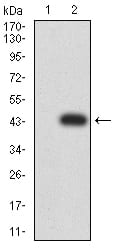
Figure 3:Western blot analysis using SYK mAb against HEK293 (1) and SYK (AA: 217-356)-hIgGFc transfected HEK293 (2) cell lysate.
Immunofluorescence analysis
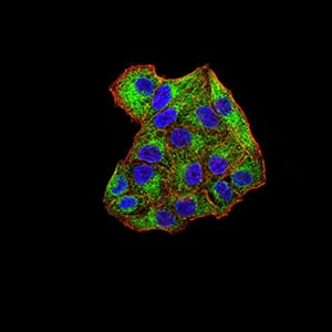
Figure 4:Immunofluorescence analysis of Hela cells using SYK mouse mAb (green). Blue: DRAQ5 fluorescent DNA dye. Red: Actin filaments have been labeled with Alexa Fluor- 555 phalloidin. Secondary antibody from Fisher (Cat#: 35503)
Flow cytometric
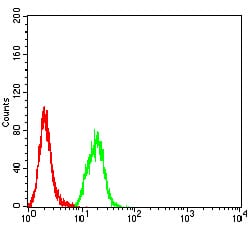
Figure 5:Flow cytometric analysis of Hela cells using SYK mouse mAb (green) and negative control (red).
For Research Use Only. Not for use in diagnostic procedures.