

SV2C Primary Antibody
Item Information
Catalog #
Size
Price
Description
SV2C (Synaptic Vesicle Glycoprotein 2C) is a Protein Coding gene. Diseases associated with SV2C include Foodborne Botulism and Alcohol-Related Birth Defect. Among its related pathways are Toxicity of botulinum toxin type F (BoNT/F) and Uptake and actions of bacterial toxins. Gene Ontology (GO) annotations related to this gene include transporter activity and transmembrane transporter activity. An important paralog of this gene is SV2A.
Product Overview
Entrez GenelD
22987
Clone#
5G11D10
Host / Isotype
Mouse / Mouse IgG1
Species Reactivity
Human
Immunogen
Purified recombinant fragment of human SV2C (AA: extra mix) expressed in E. Coli.
Formulation
Purified antibody in PBS with 0.05% sodium azide
Storage
Store at 4°C short term. Aliquot and store at -20°C long term. Avoid freeze/thaw cycles.
Product Applications
WB (Western Blot)
1/500 - 1/2000
IHC_P(Immunohistochemistry)
1/200 - 1/1000
ICC (Immunocytochemistry)
1/200 - 1/1000
FCM (Flow Cytometry)
1/200 - 1/400
ELISA
1/10000
References
1,Nat Struct Mol Biol . 2016 Jul;23(7):656-62.
2,Trends Biochem Sci . 2014 Nov;39(11):517-26.
2,Trends Biochem Sci . 2014 Nov;39(11):517-26.
Product Image
Elisa

Figure 1:Black line: Control Antigen (100 ng);Purple line: Antigen (10ng); Blue line: Antigen (50 ng); Red line:Antigen (100 ng)
Western Blot

Figure 2:Western blot analysis using SV2C mAb against human SV2C (AA: extra mix) recombinant protein. (Expected MW is 22.7 kDa)
Western Blot

Figure 3:Western blot analysis using SV2C mAb against HEK293-6e (1) and SV2C (AA: extra mix)-hIgGFc transfected HEK2936e (2) cell lysate.
Immunohistochemical analysis

Figure 4:Immunofluorescence analysis of Hela cells using SV2C mouse mAb (green). Blue: DRAQ5 fluorescent DNA dye. Red: Actin filaments have been labeled with Alexa Fluor- 555 phalloidin. Secondary antibody from Fisher (Cat#: 35503)
Immunofluorescence analysis

Figure 5:Flow cytometric analysis of Raji cells using SV2C mouse mAb (green) and negative control (red).
Immunohistochemical analysis

Figure 6:Immunohistochemical analysis of paraffin-embedded liver cancer tissues using SV2C mouse mAb with DAB staining.
Immunohistochemical analysis
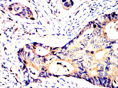
Figure 7:Immunohistochemical analysis of paraffin-embedded rectal cancer tissues using SV2C mouse mAb with DAB staining.
For Research Use Only. Not for use in diagnostic procedures.